39 diagram of the rectum
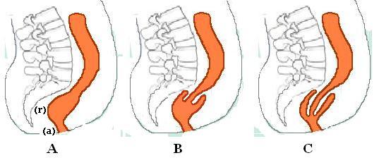
The colon is designed with squatting in mind. Among other things, it has natural built-in obstacles in the form of kinks and bends. One is a bend at the rectum which is released only when a person squats. The other bend is the kink where the sigmoid colon joins to the rectum. To release this kink, the sigmoid colon must be lifted up. Anatomy Blood supply to colon from superior and inferior mesenteric arteries Junction—relatively poor blood supply Rectum—three sources—IMA, internal iliac, internal pudendal Venous drainage of rectum to IMV/portal and to systemic circulation Connected by venous cushions--rrhoids
Mar 19, 2015 · Rectum. The rectum is the concluding part of the large intestine that terminates in the anus. The average length of the human rectum may range between 10 and 15 cm. Its diameter can be compared to ...

Diagram of the rectum
The anal canal is located within the anal triangle of the perineum between the right and left ischioanal fossae. It is the final segment of the gastrointestinal tract, around 4cm in length. The canal begins as a continuation of the rectum and passes inferoposteriorly to terminate at the anus. Anatomical Structure. The rectum is a chamber that begins at the end of the large intestine, immediately following the sigmoid colon, and ends at the anus (see also Overview of the Anus and Rectum Overview of the Anus and Rectum The anus is the opening at the end of the digestive tract where stool leaves the body. The rectum is the section of the digestive tract above the anus where stool is held before it passes ... The next prostate diagram or prostate picture shows all the parts of the amazing male sexual and reproductive functions. This last prostate image shows the back view of the prostate gland. This perspective reveals the ampulla of the ductus deferens (the ductus carries sperm from the testes-balls to the prostate gland, adding semen from the ...
Diagram of the rectum. Muscles in the abdominal wall contract, which increases pressure in the rectum and helps to push stool down. A person uses their pelvic oor muscles and the anal sphincter to control when stools are pushed out. The puborectalis muscle is a loop of muscle that wraps around the lower rectum. This muscle relaxes and allows the rectum to straighten. The anus is the opening where the gastrointestinal tract ends and exits the body. The anus starts at the bottom of the rectum, the last portion of the colon (large intestine). The anorectal line ... Structure Function in Digestion mouth 11. epiglottis 10. esophagus stomach 8. small intestine large intestine rectum 6. anus liver gall bladder pancreas 1.) Label the diagram of the digestive system with the structures given in the table to the right 2.) In the table, give the function of each of the structures you have We are pleased to provide you with the picture named Anatomical Location Of Vagina, G-spot, Clitoris, Anus.We hope this picture Anatomical Location Of Vagina, G-spot, Clitoris, Anus can help you study and research. for more anatomy content please follow us and visit our website: www.anatomynote.com. Anatomynote.com found Anatomical Location Of Vagina, G-spot, Clitoris, Anus from plenty of ...
The large bowel is made up of the colon, rectum and anus. Diagram of the digestive system. When you swallow food, it passes down the gullet (oesophagus) to the stomach. This is where digestion begins. The food then enters the small bowel, where nutrients and minerals from food are absorbed. The digested food then moves into the colon. Sigmoid colon: This is the last part of the colon. Rectum: The stool goes through the rectum. The rectum ends at the anus. Anus: This is the opening at the end of the colon. Stool leaves the body through this opening. The rectum and anus have muscles and nerves that control bowel movements. Blood supply to the colon The diagram below shows the structure and functions of the human digestive system. Let learn the different parts of the human digestive system. Mouth — It includes teeth, salivary glands and tongue. It is the beginning of the digestive tract and the process of digestion begins from the mouth, where teeth help by breaking and grinding the food ... File:Stomach colon rectum diagram-en.svg. Size of this PNG preview of this SVG file: 512 × 529 pixels. Other resolutions: 232 × 240 pixels | 465 × 480 pixels | 581 × 600 pixels | 743 × 768 pixels | 991 × 1,024 pixels.
rectum, terminal segment of the digestive system in which feces accumulate just prior to discharge. The rectum is continuous with the sigmoid colon and extends 13 to 15 cm (5 to 6 inches) to the anus.A muscular sheet called the pelvic diaphragm runs perpendicular to the juncture of the rectum and anal canal and maintains a constriction between these two segments of the large intestine. In the conic section, the latus rectum is the chord through the focus, and parallel to the directrix. The word latus rectum is derived from the Latin word "latus" which means "side", and the "rectum" which means "straight". Half the latus rectum is called the semi latus rectum. The diagram above shows the latus rectum of a parabola. (descending colon) Rectum Anus Large intestine Large Intestine In the Large Intestine: Indigestible parts of food move from the small intestine to the large intestine. Water and vitamins are absorbed back in the blood to be reused. The remaining waste passes to the RECTUM where peristalsis forces it through the ANUS and out of the body. As you look at the elementary canal diagram and observes it from anterior to the posterior, rectum forms the second last part and anus comes at the end. In other words, the rectum is the concluding segment of the digestive tract which ends at the anus. Fig. 2: Anus and Rectum diagram. Rectal region is a bit large in size as compared with the anus.
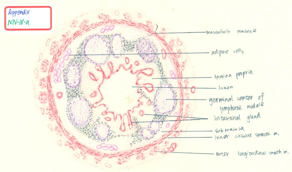
Histology Handmade Diagrams Cards for 2nd Year MBBS
The rectum is the most distal segment of the large intestine, and has an important role as a temporary store of faeces.. It is continuous proximally with the sigmoid colon, and terminates into the anal canal.. In this article we will discuss the anatomy of the rectum - its structure, anatomical relationships, and clinical relevance.

Analingus Venn Diagram
The diagram accustomed beneath represents the latus rectum of a parabola. (image will be uploaded soon).. Our video diagrams the genitalia that accomplish up the rectum and anus, and their functions. While accepting a bowel movement, the rectum contracts, stool is pushed through an aperture alleged the anus, and.

Biology 122 > Mikula > Flashcards > Digestive System ...
The anus is the final part of the gastrointestinal tract, and directly continues from the rectum.The anus passes through the pelvic floor.The anus is surrounded by muscles. The top and bottom of the anus are surrounded by the internal and external anal sphincters, two muscular rings which control defecation.: 397 The anus is surrounded in its length by folds called anal valves, which converge ...

.:Arashi:. Original Species (Censored Version)
The rectum is the final segment of the large intestine that connects the colon to the anus. It stores fecal matter produced in the colon until the body is ready to eliminate the waste through the process of defecation. Anatomy. The rectum is a hollow muscular tube about 8 inches (20 cm) in length and 2.5 inches in diameter at its widest point.

Image from page 188 of "Elements of biology, with special reference to their rôle in the lives of animals" (1933)
The anus is the opening to the lower gastrointestinal (GI) tract and connects to the rectum, which connects to the colon, which traveling backwards connects to the small intestine, then the stomach, then the esophagus and finally the mouth. The anus is approximately 2 to 3 inches long and composed of skin type cells also known as squamous cells.

Image from page 521 of "Studies on hypertrophy and cancer of the prostate" (1906)
The diagram of the digestive system that is provided in the article will give one a better understanding of this organ system, as the food moves down from the mouth, through the esophagus to the stomach, small intestine and the large intestine, before it is excreted through the rectum and the anus. The table, given below, gives a consolidated ...
File:Internal rectal intussusception.jpg
The tissues around the anus become tender and painful. On the other hand, in case of perianal abscess, the victim develops red-colored, warm and painful boil-like swelling around the anus. Whereas, as the name suggests, anorectal abscesses involve the build-up of pus in both the anus and rectum. The treatment involves the use of antibiotics and ...

File:Stomach colon rectum diagram-bg.svg
Anatomy of Colon and Rectum. The entire colon is about 5 feet (150 cm) long, and is divided into five major segments. The rectum is the last anatomic segment before the anus.. The ascending and descending colon are supported by peritoneal folds called mesentery.. The right colon consists of the cecum, ascending colon, hepatic flexure and the right half of the transverse colon.

File:Anorectum-tr.svg
The colon is the largest part of the large intestine, extending from the cecum to the rectum.It is 5 feet long and its function is to reabsorb water from digested food and concentrate solid waste material, known as stool. The colon is made of several sections. The ascending colon travels up the right side of the abdomen. The part that sits across the abdomen is called the transverse colon.

File:Stomach colon rectum diagram-af.svg
The rectum is a part of the lower gastrointestinal tract.The rectum is a continuation of the sigmoid colon, and connects to the anus.The rectum follows the shape of the sacrum and ends in an expanded section called an ampulla where feces is stored before its release via the anal canal.An ampulla (from Latin bottle) is a cavity, or the dilated end of a duct, shaped like a Roman ampulla.

Image from page 246 of "The hydropathic encyclopedia: a system of hydropathy and hygiene .." (1877)
the rectum until you are ready to have a bowel movement. In fact, squeezing the external sphincter muscle pushes the stool out of the anal canal (5) and the rectum relaxes. The urge to have a bowel movement is gone until the next colon contraction hits the rectum. Frequent holding of stools can cause constipation and desensitization of nerve cells.

Lab 1 Rat Dissection Flashcards at Sacred Heart ...
• The sigmoid colon is a short curving of the colon, just before the rectum. The colon removes water, salt, and some nutrients forming stool. Muscles line the colon's walls, squeezing its ...

Image from page 471 of "Treatise on gynaecology : medical and surgical" (1894)
It is the size of a walnut and anatomically sits in front of the rectum and between the bladder and the penis. The prostate surrounds the urethra. Sperm are generated in the testes. Tubes called vasa deferens are conduits for sperm that move from the testes to the seminal vesicles. Seminal vesicles secrete fluid that is a component of semen.

Image from page 62 of "Ants; their structure, development and behavior" (1910)
Now, the anus itself is composed of two sphincters. As you might recall from our discussion on the esophagus, sphincters are just muscles that close off a hole. There are two sphincters that are responsible for the anus. There's going to be the internal anal sphincter, and there's going to be the external anal sphincter.

Barium enema - Mayo Clinic
The large intestine is divided into the cecum, colon, rectum and anal canal.The large intestine begins at the cecum. The ileum (small intestine) ends where it connects to the cecum at the ileocecal junction.. The colon is divided into four parts: the ascending, transverse, descending and sigmoid.The ascending and transverse colon meet at the right hepatic flexure (near the liver).

File:Anorectum-zh.svg
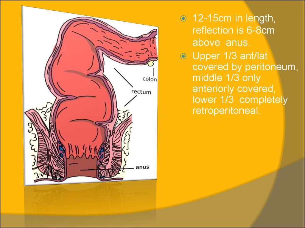
The rectum is the last part of the large intestine and connects the sigmoid colon to the anal canal.. The rectum begins at the height of S2-S3 and ends at the perineum. It is about 12 to 16 cm long und may be subdivided into three parts:. The upper third lies intraperitoneally; The middle third retroperitoneally; The lower third under the pelvic diaphragm and therefore extraperitoneally.

File:Rectum anatomy en.svg
The next prostate diagram or prostate picture shows all the parts of the amazing male sexual and reproductive functions. This last prostate image shows the back view of the prostate gland. This perspective reveals the ampulla of the ductus deferens (the ductus carries sperm from the testes-balls to the prostate gland, adding semen from the ...
Large intestine
The rectum is a chamber that begins at the end of the large intestine, immediately following the sigmoid colon, and ends at the anus (see also Overview of the Anus and Rectum Overview of the Anus and Rectum The anus is the opening at the end of the digestive tract where stool leaves the body. The rectum is the section of the digestive tract above the anus where stool is held before it passes ...

Image from page 387 of "Diseases of the ovaries : their diagnosis and treatment" (1865)
The anal canal is located within the anal triangle of the perineum between the right and left ischioanal fossae. It is the final segment of the gastrointestinal tract, around 4cm in length. The canal begins as a continuation of the rectum and passes inferoposteriorly to terminate at the anus. Anatomical Structure.

Image from page 246 of "The hydropathic encyclopedia : a system of hydropathy and hygiene in eight parts ... designed as a guide to families and students, and a text-book for physicians" (1853)

Image from page 140 of "Porneiopathology : a popular treatise on venereal and other diseases of the male and female genital system : with remarks on impotence, onanism, sterility, piles, and gravel, and prescriptions for their treatment" (1844)

Image from page 334 of "Diseases of women. A clinical guide to their diagnosis and treatment" (1899)

Image from page 435 of "The elements of Embryology" (1889)

File:Stomach colon rectum diagram-es.svg

Image from page 186 of "Elements of biology, with special reference to their rôle in the lives of animals" (1933)
Inflammatory diseases of rectum - online presentation

Image from page 105 of "Practical anatomy of the rabbit : an elementary laboratory textbook in mammalian anatomy" (1921)

Digestive at University of Tulsa - StudyBlue

Internal Anatomy of Larry Koopa

Image from page 64 of "The practice of surgery" (1910)
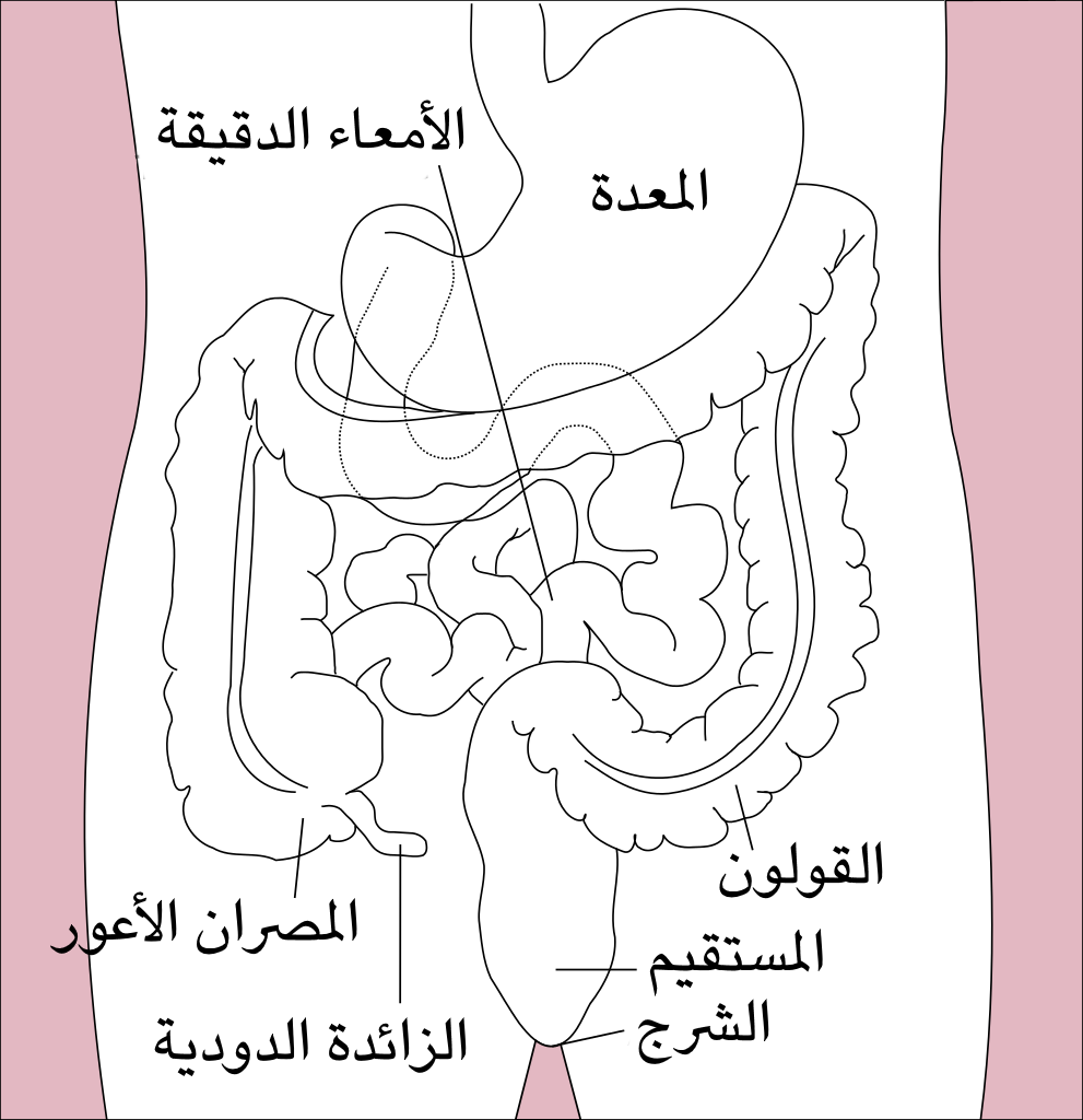
File:Stomach, colon and rectum diagram (Arabic).png

Image from page 332 of "Elements of the comparative anatomy of vertebrates" (1886)

Image from page 682 of "The chordates" (1950)

Head of Tyrannosaurus - Anatomy Primer

Image from page 890 of "Anatomy, descriptive and surgical" (1887)

File:Stomach colon rectum diagram-cy.svg

Histology Handmade Diagrams Cards for 2nd Year MBBS

Image from page 435 of "The elements of embryology" (1883)

File:Stomach colon rectum diagram-ta.png
0 Response to "39 diagram of the rectum"
Post a Comment