40 growth plate in wrist diagram
Answer (1 of 2): According to this diagram, the last to fuse was sacrum. I really never bother to memorize the sequence. There is other diagram depicting the growth plate fusion on paediatric patient. The first one to fuse is bone in finger. There is also a drawing about the bone in hand. B... A growth plate injury is an injury to the growth plates, which are located on each end of long bones. Children and teens with growth plate injuries often need immediate treatment to prevent problems with bone growth. Depending on the type of injury, your child may need surgery and a cast or splint.
Grade 4A actually looks more fused than Grade 3A. "Grade 5 growth plate. (A) There is less than 5 mm fusion left of the fusion of the growth plate in plain radiograph. (B) Similar correlating appearance on T1-weighted MRI of the same patient (arrows)."<-And 5A looks even less fused. 5B however does look more fused than 4B. "Grade 6 growth plate.
Growth plate in wrist diagram
The pubertal growth spurt is a vital period in the orthodontic treatment and should be kept in mind when planning orthodontic treatment in growing children. One of the main objectives of taking hand and wrist radiograph is to determine the amount of growth and get used of it in patients with skeletal discrepancy during adolescence. Jul 15, 2019 · The growth plates in the hands and wrist are particularly susceptible to injury through falling on hands and wrists. Growth plates are weaker than the surrounding bone simply because they are not yet fully ossified. What Are Growth Plate Injuries? Growth plate injuries are usually fractures that affect the area in question. Wrist anatomy is the study of the bones, ligaments and other structures in the wrist. The wrist joint is a complex joint which connects the forearm to the hand, allowing a wide range of movement. ... A distal radial epiphysis injury is an injury to the growth plate at the wrist end of the radius bone in the forearm. It mostly…
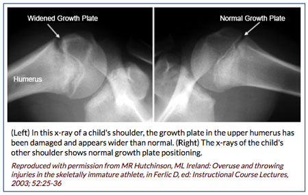
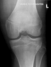
Growth plate in wrist diagram. In particular, a widening of the growth plate may be seen. If there is a narrowing of the growth plate seen on the X-ray, then a Salter-Harris type stress fracture should also be considered. Ganglion cysts, wrist tendonitis or other joint dysfunction which may also present should be ruled out. It is important not to miss a Scaphoid fracture. Hand and Wrist Growth Plate Injuries. Hand and wrist injuries are perhaps the most common injuries among active children. The growth plates in the hand and wrist are at risk of injury and fracture because the cartilage located in these areas is weaker than surrounding ligaments. While an injury to the growth plate in the wrist or hand usually heals without complication, it is important to seek treatment in a timely manner to help prevent potential long-term growth and healing problems. The growth plates around the knee are more sensitive to injury. A growth plate fracture at the knee can cause the leg to be shorter, longer or crooked if the growth plate has permanent damage. Growth plate injuries around the wrist and shoulder usually heal without problems. FPnotebook.com is a rapid access, point-of-care medical reference for primary care and emergency clinicians. Started in 1995, this collection now contains 6990 interlinked topic pages divided into a tree of 31 specialty books and 736 chapters.
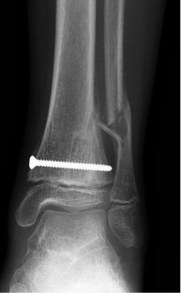
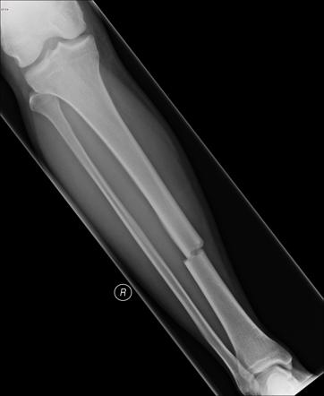
Description. Most growth plate fractures occur in the long bones of the fingers. They are also common in the outer bone of the forearm (radius) and lower bones of the leg (tibia and fibula). Growth plate fractures vary greatly with regard to risk for growth problems. Factors that affect the risk of problems over time include: The patient's age. Answered by : Dr. Grzegorz Stanko ( General Surgeon) Having chronic hives after plate and screws put in wrist after fracture. Blood work done. Worrisome. MD. On 3/16/2012 I had a plate and screws put in my wrist caused by a fracture . Pediatric Distal Forearm and Wrist Injury: An Imaging Review1 Injuries to the pediatric distal forearm and wrist have myriad mani-festations. Growth plate injuries can occur in the skeletally imma-ture child. An unfused growth plate is less robust than ligamentous complexes and therefore is more easily injured. The Salter-Harris Gymnast's Wrist is irritation and inflammation of the growth plate (epiphysis) at the end of the radius (forearm bone) where it connects to the hand to form the wrist. In a child, the bones grow from areas called growth plates. The growth plate is made up of cartilage, which is softer and more vulnerable to injury than mature bone.
Salter-Harris type II wrist growth plate injury. Gymnasts' Wrist Page 1 of 4 3.31.09 Gymnasts' Wrist - Distal Radius Growth Plate Injury Background 1. Definition o Chronic stress injury of distal radial physis o Also known as epiphysiolysis 2. General info o Common injury in pediatric gymnast o Diagnosis made by history and physical exam Confirmed with plain x-rays or MRI Treatment often nonsurgical Growth plates are areas of developing cartilage tissue near the ends of long bones in children. The growth plate helps determine the length and shape of the adult bone. As a bone grows, the shape and size of a cyst can change. The cyst will stop growing when the child is full-grown, and then will gradually fill in with normal bone and disappear ... All children have "growth plates" - areas of smooth, elastic cartilage found at the end of each long bone in the body. This is where growth takes place. When bones finish growing, the growth plates close. Girls generally stop growing and reach their maximum height between ages 14 and 16, and boys finish their growth between 16 and 18 years ...
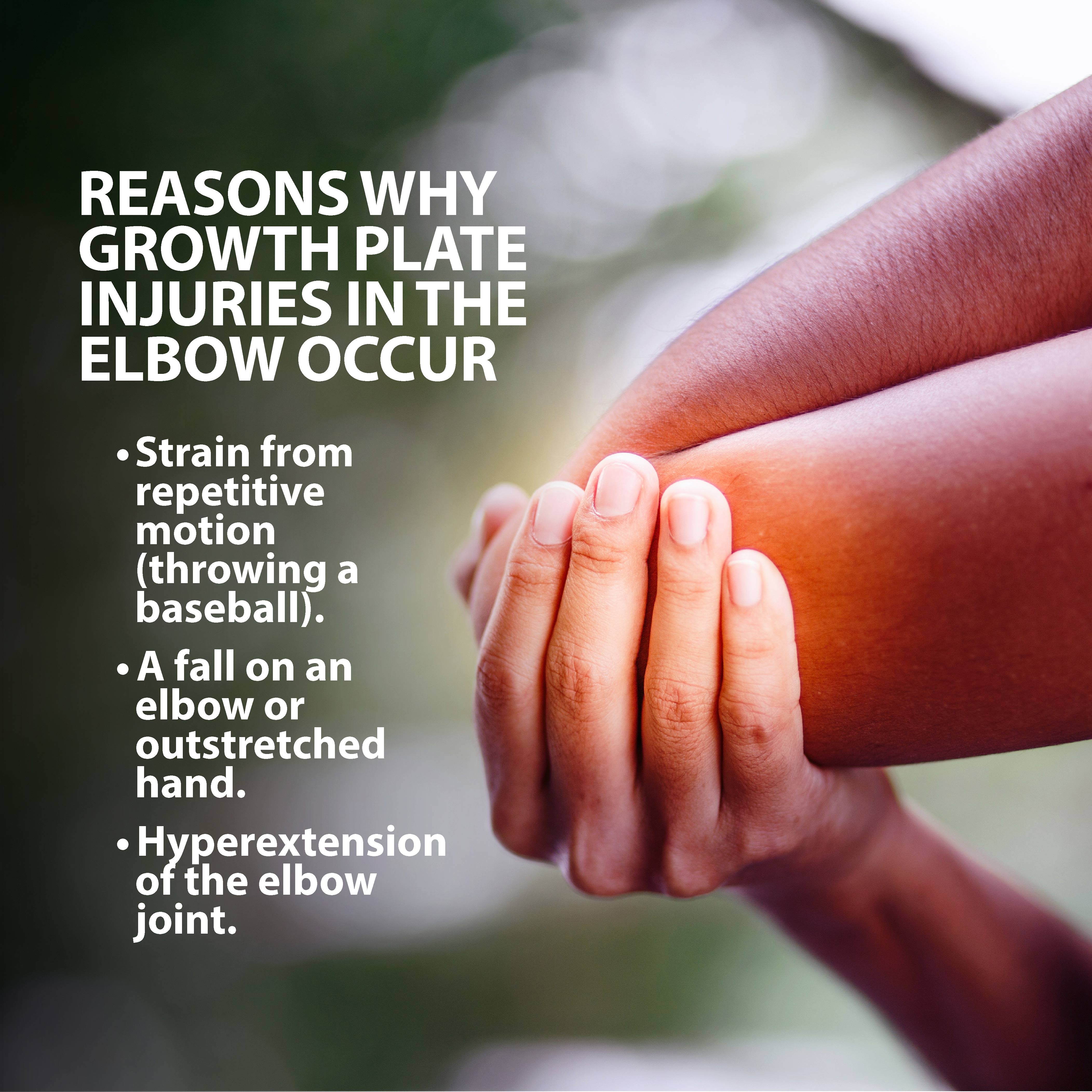
Growth Plate Injuries Of The Elbow Florida Orthopaedic Institute
Access 130+ million publications and connect with 20+ million researchers. Join for free and gain visibility by uploading your research.
Salter Harris Type Ii Wrist Growth Plate Injury Radiology Case Radiopaedia Org
The growth plates in the hand and wrist are at risk of injury & fracture because the cartilage located in these areas is weaker than surrounding ligaments.
If Growth Plates On The Wrists Are Closed Does That Mean Growth Plates Everywhere Else In The Body Are Closed Quora
Learn how growth plate fractures are diagnosed and treated at Boston Children's Hospital.
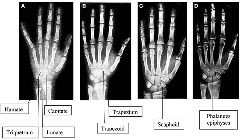
Frontiers Evaluation Of Bone Age In Children A Mini Review Pediatrics
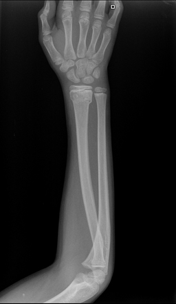
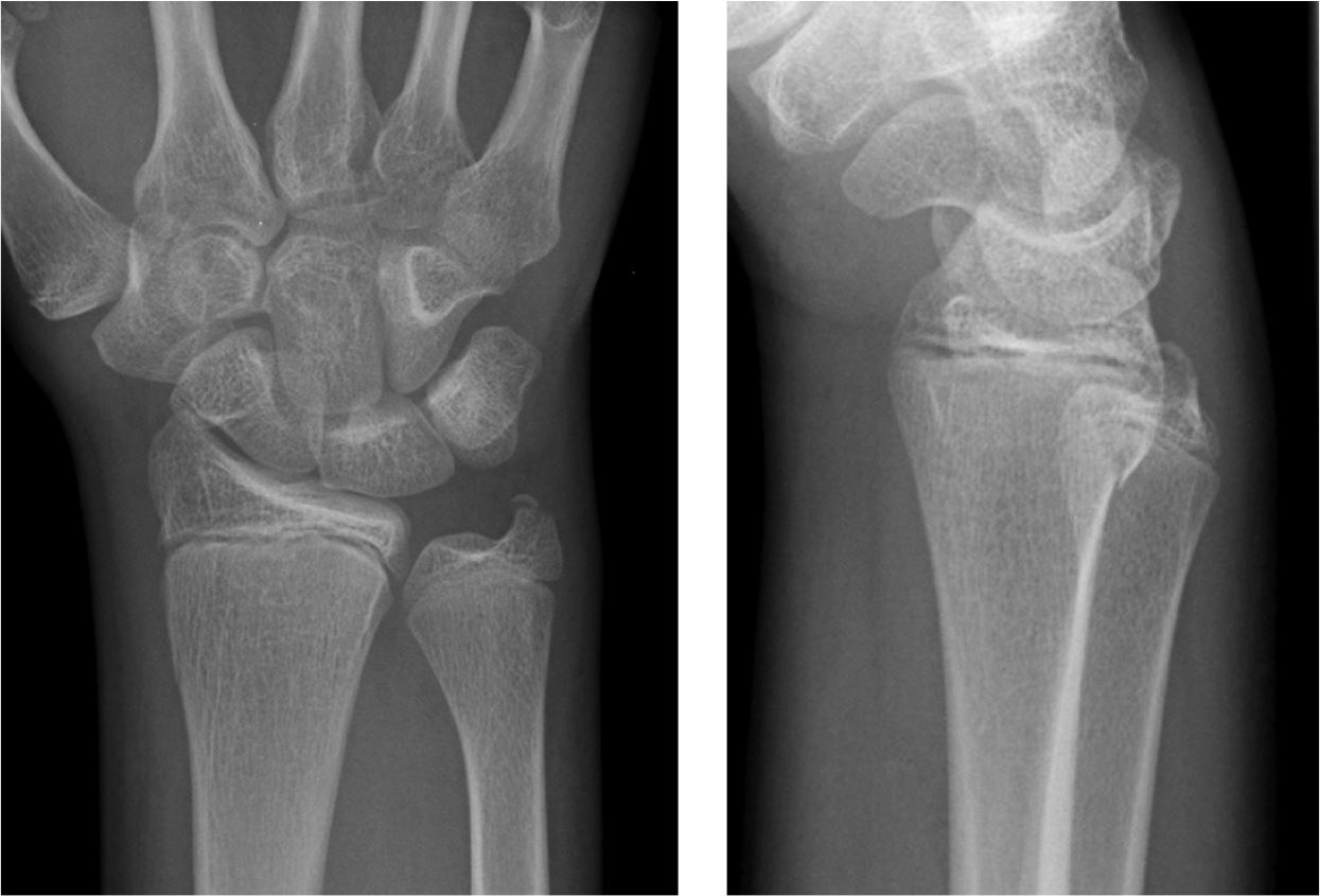
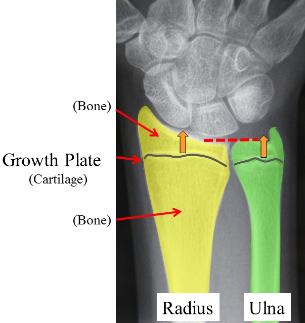
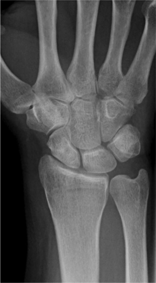
The example is of a wrist, partly because about 50% of growth plate injures are at the wrist and partly because that is the most likely growth plate injury my patients will have. In this xray, the wrist bones are at the top and the forearm bones at the bottom; the radius is the bone on the right, it is the one on the thumb side of the hand ...
Risk Injury In Young Athletes Bouldercentre For Orthopedics Spine
Injuries to the pediatric distal forearm and wrist have myriad manifestations. Growth plate injuries can occur in the skeletally immature child. An unfused growth plate is less robust than ligamentous complexes and therefore is more easily injured. The Salter-Harris fracture classification system is used to grade physeal injuries based on their imaging appearance. This grading has prognostic ...
Epiphyseal Growth Plate High Resolution Stock Photography And Images Alamy
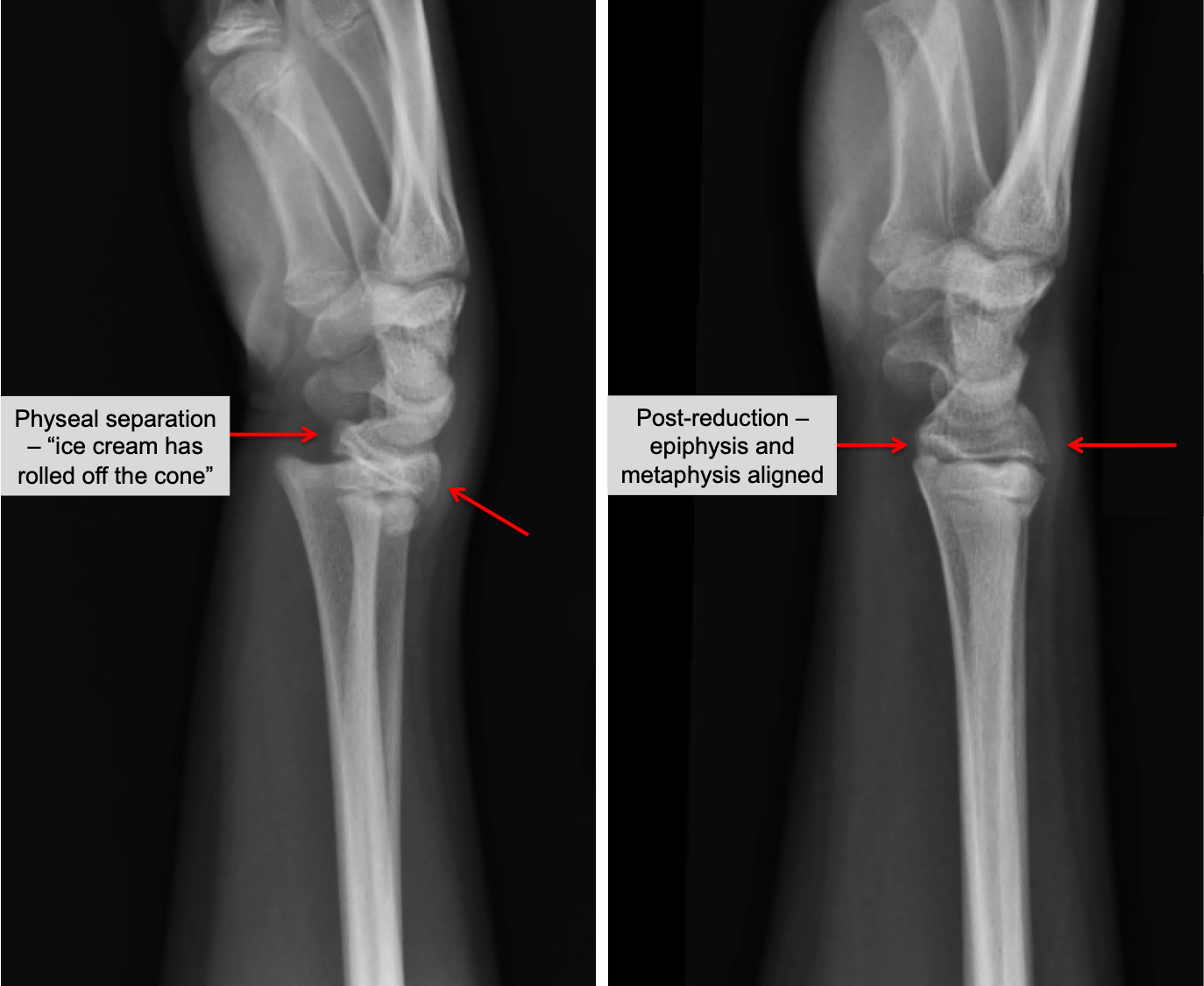
The peak age for injury to the growth plate is in the pre-adolescent growth spurt. The Salter-Harris type II fracture is the most common type. Distal radial physeal fractures are uncommon in children younger than five years. The most common mechanism of injury is a fall on an outstretched hand (Figure 1).

Longitudinal Analysis Of Pediatric Distal Radius Alignment Parameters In A Cohort Of Serial Radiographs Knapik 2021 Clinical Anatomy Wiley Online Library
Learn how to keep kids safe while still having fun · Find online support groups and forums for pediatric orthopaedic conditions

Growth Plate Fracutres
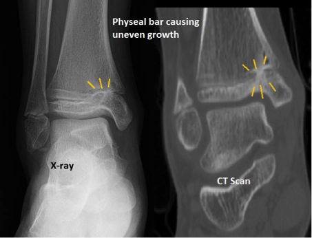
Injuries to growth plates, which produce new bone tissue and determine the final length and shape of bones in adulthood, must be treated so that bones heal properly.
Orthokids Growth Plate Physeal Fractures
November 19, 2019 - At Connecticut Orthopaedics, our hand surgeons will provide you with an accurate diagnosis and treatment plan that’s specific to you. Get started

Hand Growth Plates
Growth Plate (Physeal Fractures) Pediatric Orthopaedic Society of North America (POSNA) 1 Tower Ln, Suite 2410 Oakbrook Terrace, IL 60181 p: (630) 478-0480 f: (630) 478-0481 e: posna@posna.org Find A Pediatric Orthopaedist ...
Epiphyseal Growth Plate High Resolution Stock Photography And Images Alamy
Answer: Fusion of the growth plate (epiphysis) is most often the same for the left and right sides but can vary among the bones of the body. Forensic scientists use tables (such as the one above) to estimate the age of death from skeletal remains. (Consider what would happen if epiphyses closed ...

The Growth Plate Connecticut Orthopaedics
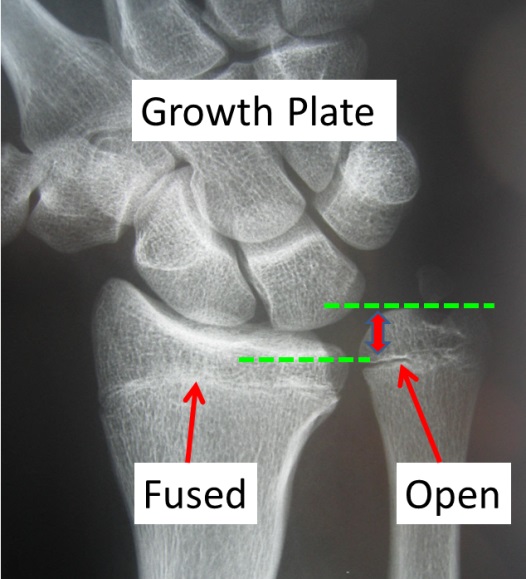
Growth plates are one way bones grow. There are usually two growth plates in each long bone. They add length and width to the bone. As kids grow, the growth plates harden into solid bone. A growth plate that has completely hardened into solid bone is a closed growth plate. After a growth plate closes, the bones are no longer growing.
The Wrist
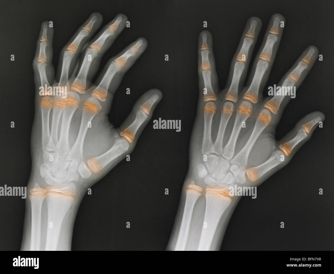
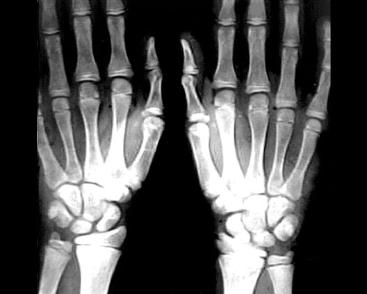
A child's bones, such as those in the fingers and wrist, contain "growing zones" at both ends called growth plates. These plates consist of special cells responsible for the bones' growth in length. Growth plates are easy to spot on an X-ray because they're softer and contain less mineral, making them appear darker on an X-ray image than the ...

Radiology Of Fracture Principles Ppt Video Online Download
Scott H. Kozin MD, in Principles and Practice of Wrist Surgery, 2010. Growth Plate Arrest. Growth plate closure occurs in approximately 4% to 5% of all Salter-Harris distal radius fractures. 21,22 Therefore, all growth plate fractures mandate a follow-up x-ray 3 to 6 months after healing to ensure continued growth.
Growth Plate Fractures Orthoinfo Aaos
August 7, 2007 - Learn about growth plate fractures and injuries in children. Causes include overuse injuries, sports, childe abuse, juvenile arthritis, frostbite, radiation, and chemotherapy. Diagnosis is based on the Salter Harris Classification system.
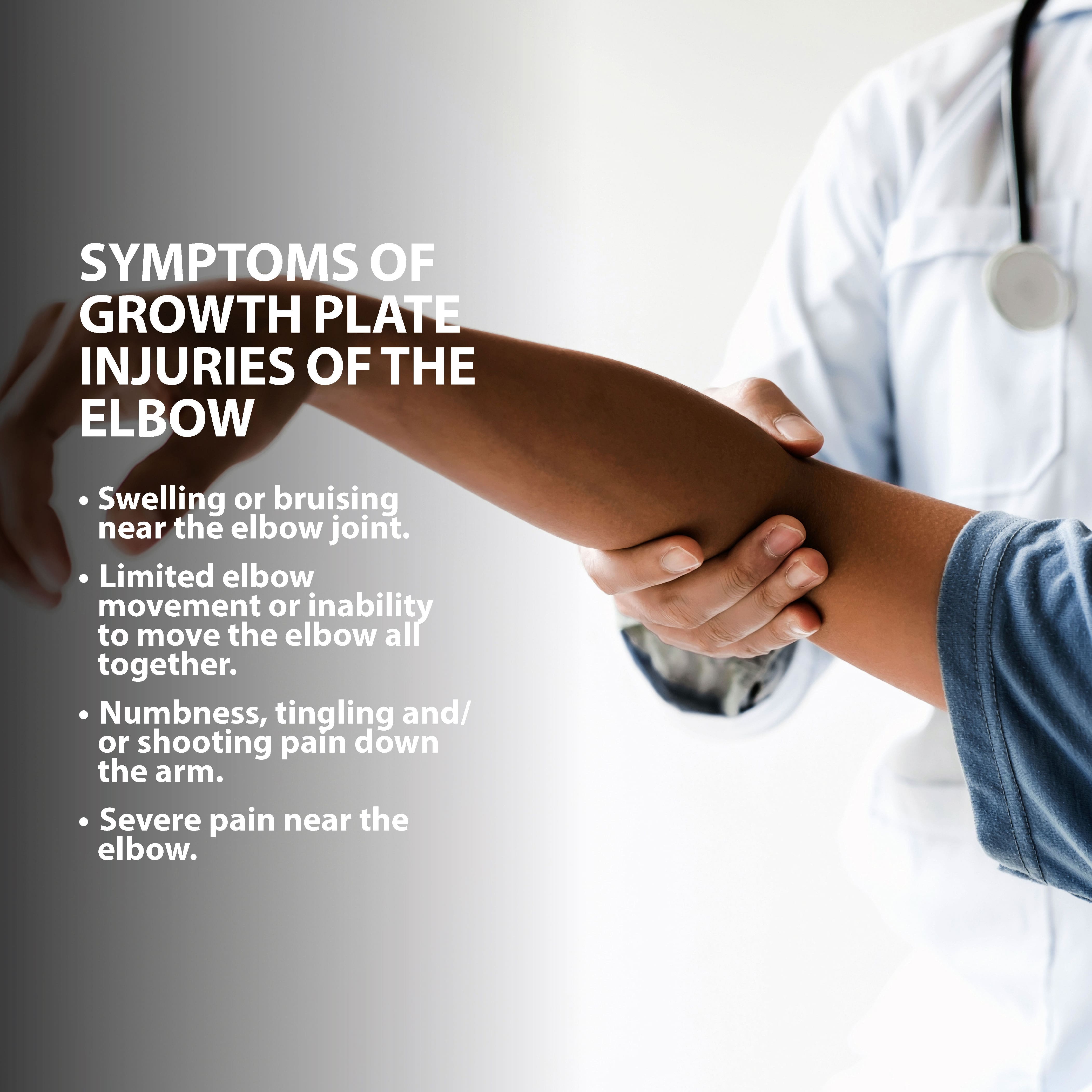
Growth Plate Injuries Of The Elbow Florida Orthopaedic Institute
Learn about Boston Children’s Hospital, ranked the #1 pediatric hospital in the country by U.S. News and World Report.
Orthokids Growth Plate Physeal Fractures
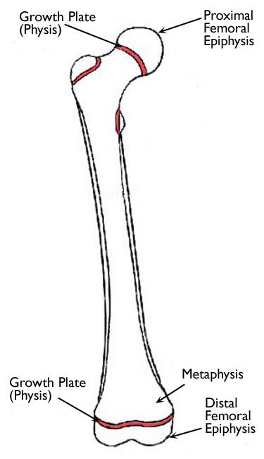
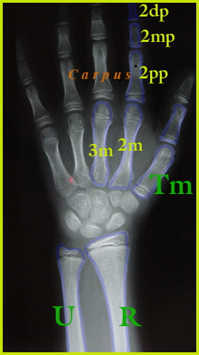
The physis appears as a radiolucent line in skeletally-immature patients located between the metaphysis and epiphysis. It contains zones of mesenchymal cells in various maturation stages (see physeal anatomy illustration). As the metaphysis and epiphysis mature and fuse, the physis thins, disappears, and endochondral ossification ceases.
Children S Wrist Fractures
As most orthodontic patients are growing individuals, orthodontists have to consider the remaining craniofacial growth of each patient for successful treatment planning. During treatment, growth can cause significant changes in the dental and skeletal structures as well as in the profile. Abundant studies have shown that such changes are complex due to the fact that each patient has an individual growth pattern (Björk, 1951, 1963; Nanda, 1955; Kraus et al., 1959; Bambha and Van Natta, 1963; Johnston et al., 1965; Mitani, 1977; Fishman, 1979; Bishara et al., 1984; Bishara and Jakobsen, 1985). Accordingly, onset duration, velocity, direction, and the amount of remaining growth differ significantly among individuals with the same chronological age. Remaining craniofacial growth can contribute to orthodontic correction as it is expected in patients with a Class II malocclusion. On the other hand, remaining growth can also have an adverse effect on a skeletal malocclusion, particularly i...

Growth Plate Fractures Part 2
Mar 21, 2019 · Injuries to the growth plates can happen with any type of injury to bones in children and are very common. The fingers, wrist and lower leg bones are where these injuries most often occur, but we see them in all long bones. Growth plate injuries can occur with a single event like a fall, accident or sports-contact injury.

Next Level Sports Medicine Growth Plate Fractures
Growth plate fractures commonly result from falls or sports, bike and motor vehicle accidents. In some cases, a growth plate may fracture due to an overuse of a joint.
Describing A Fracture An Approach Radiology Reference Article Radiopaedia Org
Answer (1 of 2): According to this diagram, the last to fuse was sacrum. I really never bother to memorize the sequence. There is other diagram depicting the growth plate fusion on paediatric patient. The first one to fuse is bone in finger. There is also a drawing about the bone in hand. B...
The Wrist
The example is of a wrist, partly because about 50% of growth plate injures are at the wrist and partly because that is the most likely growth plate injury my patients will have. In this x-ray, the wrist bones are at the top and the forearm bones at the bottom; the radius is the bone on the right, it is the one on the thumb side of the hand ...
Radiology Images
Download scientific diagram | Salter-Harris fracture classifications. ... Injuries to the pediatric distal forearm and wrist have myriad manifestations. Growth plate injuries can occur in the ...

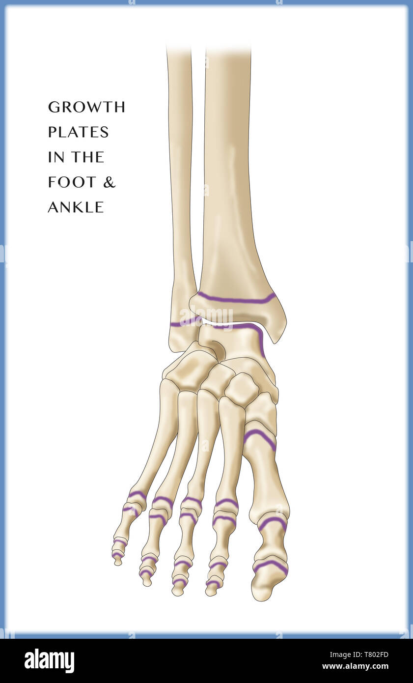
Foot Growth Plates
October 30, 2019 - When you're in the ER with your child because they broke a bone, you may hear the doctor say they need to check if it's a "growth plate" fracture. It can sound a little scary, but most injuries like that heal easily. There can be complications, though, if it's not treated correctly or if the ...

Basic Principles In The Assessment And Treatment Of
Wrist anatomy is the study of the bones, ligaments and other structures in the wrist. The wrist joint is a complex joint which connects the forearm to the hand, allowing a wide range of movement. ... A distal radial epiphysis injury is an injury to the growth plate at the wrist end of the radius bone in the forearm. It mostly…
Wrist Hand
Jul 15, 2019 · The growth plates in the hands and wrist are particularly susceptible to injury through falling on hands and wrists. Growth plates are weaker than the surrounding bone simply because they are not yet fully ossified. What Are Growth Plate Injuries? Growth plate injuries are usually fractures that affect the area in question.

Determining Age Using Bones How Bones Show Age
The pubertal growth spurt is a vital period in the orthodontic treatment and should be kept in mind when planning orthodontic treatment in growing children. One of the main objectives of taking hand and wrist radiograph is to determine the amount of growth and get used of it in patients with skeletal discrepancy during adolescence.
Pubs Rsna Org

Jaypeedigital Ebook Reader

Fracture General Principles Startradiology
Fracture Education Physeal Growth Plate Injuries
Kid S Wrist Fracture Raleigh Hand Surgery Joseph J Schreiber Md

Pediatric Hand And Wrist Fractures Hand Institute Of Charleston

Abc Of Emergency Radiology The Wrist The Bmj
1
Premature Physeal Closure Of An Extraphyseal Distal Radius Fracture Secondary To Smooth Kirschner Wire Fixation A Case Report
Wrist Fractures In Children

Epiphyseal Plate An Overview Sciencedirect Topics

Growth Plate Injuries Of The Elbow Southern California Orthopedic Institute
Medecine Uottawa Ca
0 Response to "40 growth plate in wrist diagram"
Post a Comment