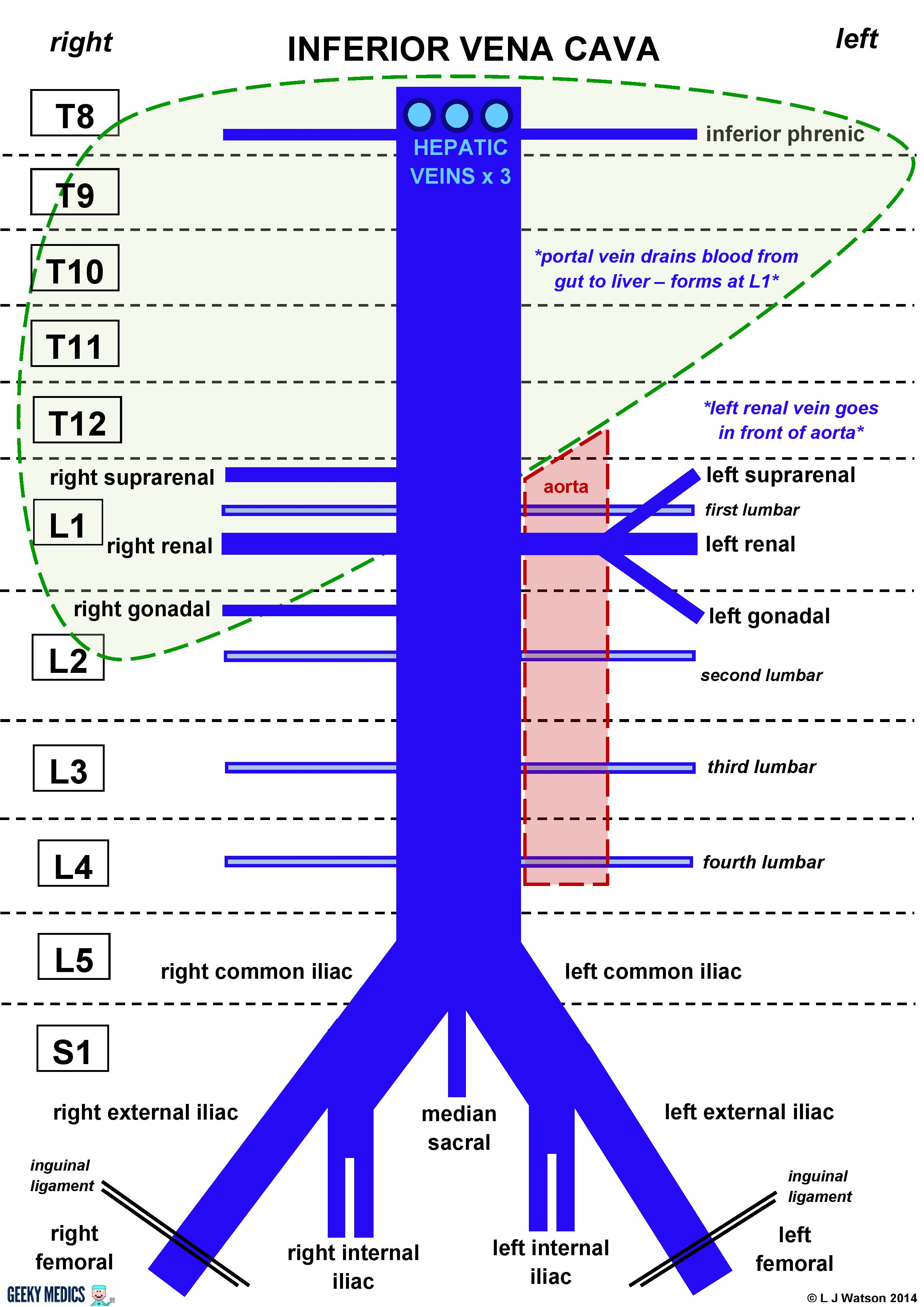
41 inferior vena cava diagram
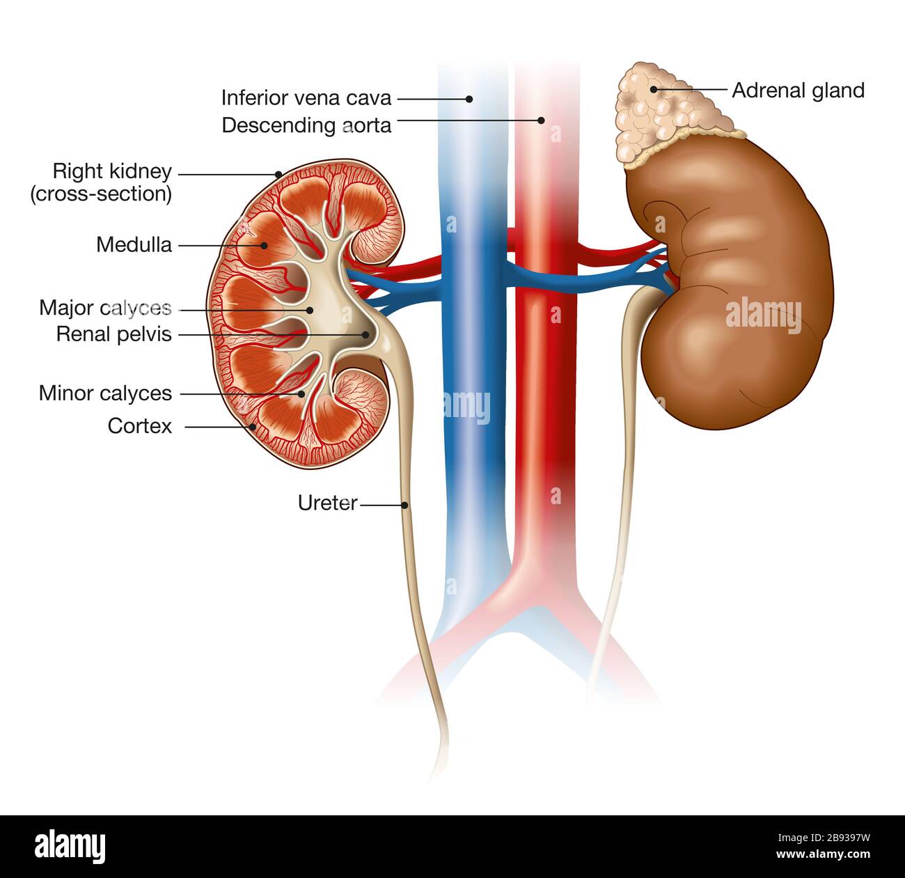
(c) Vena cava. Answer: Vena cava is the largest vein present in the body, which is divided into 2 parts such as superior vena cava and inferior vena cava. They carries blood to the heart. 3) Name any four heart related condition that occur normally in human. Briefly explain the cause and symptoms of each of them. The renal vein exits each kidney to join the inferior vena cava, which transports blood back to your heart. Kidney Function. The urinary system depends on proper kidney structure and function. Some of these core actions include: Excretes waste: The kidneys get rid of toxins, urea, andexcess salts. Urea is a nitrogen-based waste product of cell ...
RCC can invade into the renal vein and then into the inferior vena cava. From here, it can metastasize hematogenously to lung and bones. RCC can also have paraneoplastic effects. These include Cushing-like symptoms and hypercalcaemia and are caused by release of adreno-corticotropic hormone (ACTH) or parathyroid hormone-related protein (PTHrP).
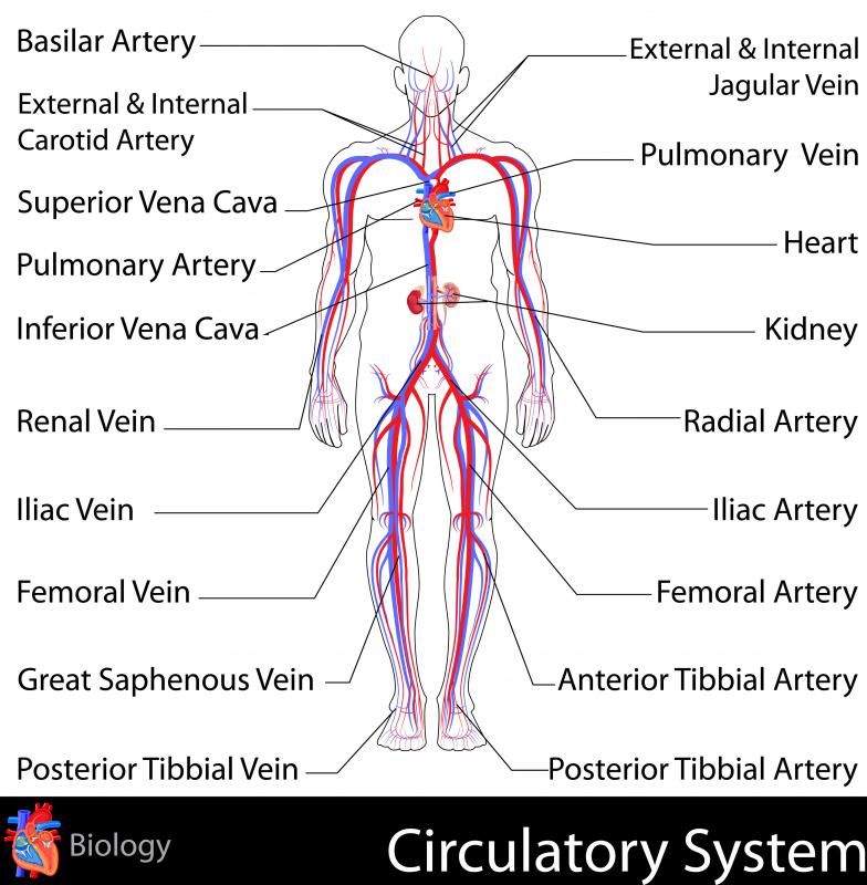
Inferior vena cava diagram
Accessory renal arteries are a common variant and are present in ~25% (range 20-30%) and are bilateral in ~10% of the population 1.Their proper identification is of utmost importance for surgical planning prior to live donor transplantation 2,3 and renal artery embolization for various reasons 4,5.. The term extra renal artery may be used 6, with a subclassification into: 6 Jul 2019 — The superior vena cava and inferior vena cava are the two largest veins in the body. Their job is to return oxygen-depleted blood to the ... The inferior vena cava (IVC) is the largest vein of the human body. It is located at the posterior abdominal wall on the right side of the aorta.Definition and function: The vein that collects d...Clinical relations: Inferior vena cava thrombosisTributaries: Inferior Phrenic, right Suprarenal, ...Source: Common iliac veins (L5)
Inferior vena cava diagram. It takes a posterior course around the superior vena cava and continues across the crista terminalis toward the Eustachian ridge (valve of the inferior vena cava). The pathway then enters the interatrial septum (above the point of the coronary sinus ) where it enters the AV node through its posterior surface. Vena Cava Superior, Aorta, Trncus pulmonalis, Venae pulmo... Anterior view of the inferior and superior vena cava in relationship to the ribcage ... Gehirn: MRT-Atlas der menschlichen Anatomie The right atrium receives the deoxygenated blood from the body. This is brought to it by the inferior and superior vena cava. The blood then flows down into the right ventricle. From here, it is sent to the lungs for oxygenation. This is done by the pulmonary artery - the only artery that carries deoxygenated blood. The inferior vena cava (IVC) (plural: inferior venae cavae) drains venous blood from the lower trunk, abdomen, pelvis and lower limbs to the right atrium of ...
Increased hydrostatic pressure is the main factor in the production of fluid in CCF, constrictive pericarditis, inferior vena cava obstruction, Budd-Chiari syndrome In the case of exudative ascites inflammatory cytokine causing increased vascular and peritoneal permeability plays a key role. 24 Feb 2020 — The inferior vena cava is also referred to as the posterior vena cava. The inferior vena cava is a large vein that carries deoxygenated ... Explore this interactive 3-D diagram to learn more about the gallbladder and gallstones. Dorsal portion sits in the oropharynx. The dilated vertical vein on the left brachiocephalic vein on top and the superior vena cava on the right form the head of the snowman. The body of the snowman is formed by the enlarged right atrium. by WD Tucker · 2020 · Cited by 4 — The inferior vena cava (IVC) is a large retroperitoneal vessel formed by the confluence of the right and left common iliac veins.
The caval opening (cavus hiatus), through which the inferior vena cava and parts of the phrenic nerve travel; In addition to these openings, several smaller openings also allow smaller nerves and blood vessels to run through. Location . The diaphragm spans across the body from the front to the back. It is the floor of the thoracic cavity and ... SUMMARY 1. Deoxygenated blood enters right atrium through Superior and Inferior Vena Cava 2. Blood enters right ventricle through tricuspid valve 3. Blood ... We Have got 7 pix about Blood Flow Diagram images, photos, pictures, backgrounds, and more. In such page, we additionally have number of images out there. The mediastinum is the thoracic area between the 2 pleural cavities. The mediastinum contains vital structures of the circulatory, respiratory, digestive, and nervous systems including the heart and esophagus, and major thoracic vessels including the superior vena cava, inferior vena cava, pulmonary arteries, pulmonary veins, and aorta. Variant anatomy. Variation in hepatic arterial anatomy is seen in 40-45% of people. Classic branching of the common hepatic artery from the celiac artery, and the proper hepatic artery into right and left hepatic arteries to supply the entire liver, is seen in only 55-60%. A single or double cystic artery may arise off the proper hepatic artery.
The correlation between intraoperative inferior vena cava-collapsibility index (IVC-CI) under TEE and central venous pressure (CVP) will also be explored. Discussion This study is the first prospective randomized clinical trial to determine the effect of low versus standard PP on gas embolism using TEE during elective LLR.
Hey meddit, I hope this is the right place to ask this question. I'm trying to do some research on the tissue surrounding the inferior vena cava and abdominal Aorta (just prior to the iliac bifurcation/about kidney level). I can't seem to find any information other than diagrams that detail it as adipose tissue. Does anyone know any additional info, or maybe a name for the region beyond retroperitoneal. Thanks for your help!
The superior and inferior mesenteric veins, draining the jejunum down as far as the upper rectum, along with the splenic vein draining the spleen, pancreas, and stomach, unite to form the hepatic portal vein, which empties blood into the liver. Toxins are filtered out by the liver, and the filtered blood is returned to the inferior vena cava ...
5202009 110716 PM. Place the pathway of blood through the heart in the correct sequence. Account Suspended Heart Diagram Human Heart Diagram Biology Worksheet Label-the-heart-answers 16 Downloaded from devkubotastorepl on November 30 2021 by guest Kindle File Format Label The Heart Answers Thank you extremely much for downloading label the heart answersMost likely you have...
A journey through the circulatory system. Another adventure with Richard Harrison… A journey through the circulatory system. Richard Harrison, inventor of the one and only shrinkinator, has long since upgraded the shrinkinator’s shrinking effects. Lasting up to 48 hours and can shrink down to the size of a singleerythrocyte.(Red blood cell) And not only that, there is a regrowth delay, that lasts about 2 hours in case of any erhm, certain emergencies. So no more RIP test subjects. Speaking of ...
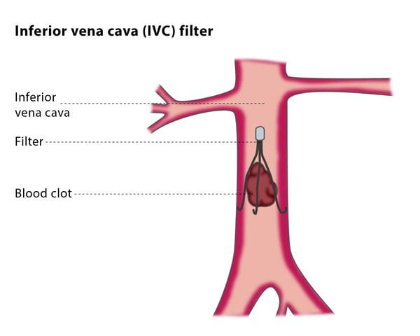
The inferior vena cava is the common convergence of venous drainage from all structures below the diaphragm. It is located on the posterior abdominal wall; anteriorly to the vertebral column and to the right of the abdominal aorta. The vessel is formed by the union of the common iliac veins at the L5 vertebral level.
Hey Reddit! I made this real quick because I'm trying to study for finals and I found this great practice questions online but with no key answer sheets. Could you guys help me? Ps, Im doing these questions to get familiar with the exam and I have picked my answers but I'm not sure if they are the correct answers. Please help me find the right answer. Thank you! BIOL 2402 practice questions 1. The blood vessels that feed directly into the capillary beds are called ________. A) muscular ar...
The inferior vena cava is a large vein that carries the deoxygenated blood from the lower and middle body into the right atrium of the heart.Drains to: Right atriumLatin: vena cava inferiorArtery: abdominal aortaAcronym(s): IVCStructure · Clinical significance
**Other discussions** [Episode 1 - Pneumococcus](https://reddit.com/r/anime/comments/8z6vpb/hataraku_saibou_ep_1_doctors_notes/) [Episode 2 - Scrape wound](https://www.reddit.com/r/anime/comments/90bxnl/hataraku_saibou_ep_2_doctors_notes/) [Episode 3 - Influenza](https://reddit.com/r/anime/comments/913mov/hataraku_saibou_ep_3_doctors_notes/) [Episode 4 - Food poisoning](https://reddit.com/r/anime/comments/93a8xt/hataraku_saibou_ep_4_doctors_notes/) [Episode 5 - Cedar pollen allergy](https:/...
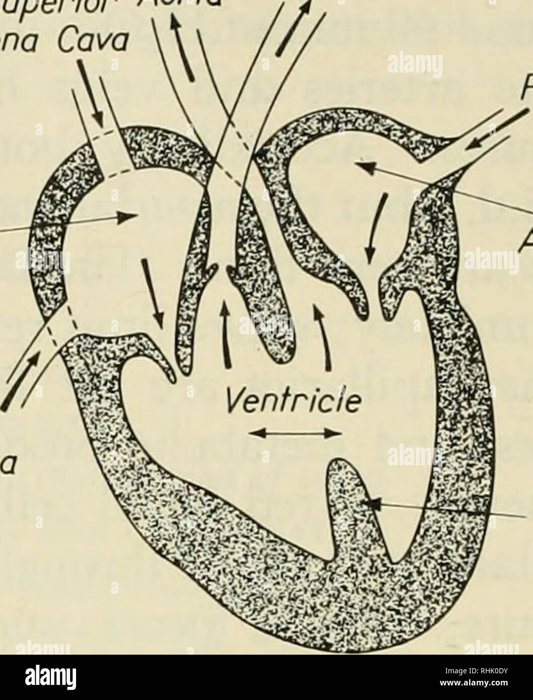
The oxygenated blood from the left ventricle is pumped to the body through the aorta and its branches. The deoxygenated blood returns to the right atrium through two large veins called the superior vena cava from the upper part of the body and the inferior vena cava from the lower part of the body.
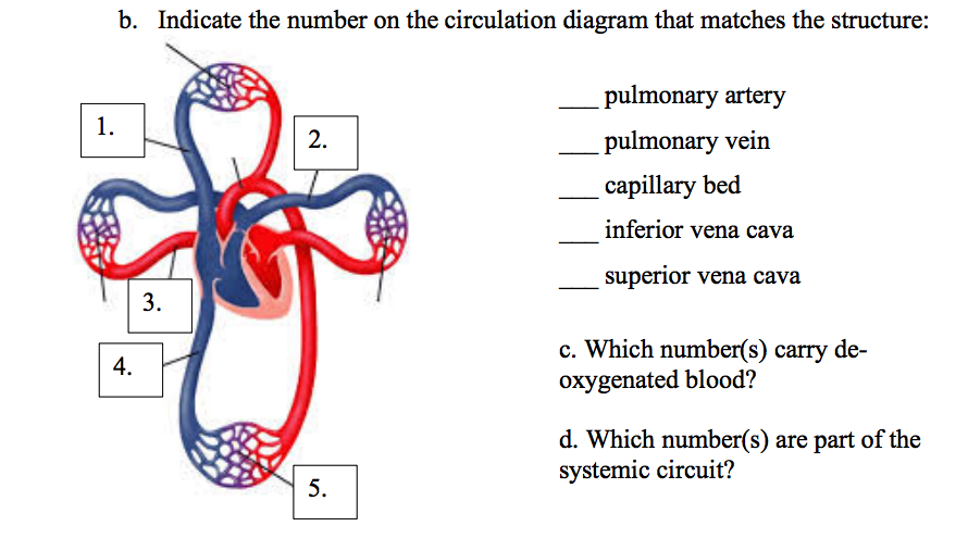
Inferior vena cava. Superior vena cava. Refer to the diagram above of the cardiovascular system and explain how blood flows through the heart. Describe the functions of the cardiovascular system. 2. Draw an arrow to identify the parts of the respiratory system to their location on the diagram.
Blood returns to the heart from the body via two large blood vessels called the superior vena cava and the inferior vena cava. This blood carries little oxygen, as it is returning from the body where oxygen was used. The vena cavas pump blood into the right atrium and the cycle of oxygenation and transport begins all over again.
Answer: Before you are born, you have a hole in your heart to bypass some of the lung circulation (not all). Most of the time, this hole (Foramen Ovale) closes after birth with lung expansion. Rarely it doesn't and NOTHING HAPPENS … the baby is fine because all it has to do is eat and poop. Later...
The inferior vena cava is a large, valveless, venous trunk that receives blood from the legs, the back, and the walls and contents of the abdomen and pelvis ...
Right atrium left atrium right ventricle left ventricle aorta caudal vena cava pulmonary artery coronary artery cranial vena cava. The worksheets should take approximately 10-15 minutes each to complete and pupils will be able to work independently through the activities. ... Right left superior and inferior. Heart Diagram Answer Keyindd Author ...
The aorta is about an inch wide and supplies oxygen-rich blood to the body's cells. The used blood them moves back and is collected into the two veins, the superior vena cava that receives blood from your upper body, and the inferior vena cava that receives blood from your lower body.
The serous pericardium is thin and consists of two parts: 1) The parietal layer lines the inner surface of the fibrous. 2) The visceral layer adheres to the heart and forms its outer covering. The parietal and visceral layers of serous pericardium are continuous at the roots of the great vessels. The narrow space created between the two layers ...
Your heart circulatory system. Label and color the superior vena cava blue. Click on the coloring page to open in a new window and print. Label and color the inferior vena cava blue. Today we are going to learn about the anatomy structure of organisms and their parts. Free printable anatomy coloring pages for kids and adults.
The upper portion of the vena cava is known as the superior vena cava, and the lower portion of the vena cava is known as the inferior vena cava. Blood from the upper portion of the body passes ...
inferior vena cava: A large vein that carries oxygen-poor blood to the right atrium from the lower half of the body. left atrium: The left upper chamber of the heart. It receives oxygen-rich blood from the lungs via the pulmonary vein. left ventricle: The left lower chamber of the heart. It pumps the blood through the aortic valve into the aorta.
[Case 1](http://www.reddit.com/r/nosleep/comments/2qkcl7/an_unknown_roundworm/) | [Case 2](http://www.reddit.com/r/nosleep/comments/2qnm72/case_2_the_eyes_in_the_sun/) | [Case 3](http://www.reddit.com/r/nosleep/comments/2qshqx/case_3_a_foreign_object_in_the_brain/) | [Case 4](http://www.reddit.com/r/nosleep/comments/2r2jh4/case_4_hypersexuality_following_parasitosis/) | [Case 5](http://www.reddit.com/r/nosleep/comments/2rd597/case_5_a_fatal_envenomation/) | [Case 6](http://www.reddit.com/r/nosle...
Interpretation: Volume status based on IVC alone (Respirophasic IVC Variation) Inferior vena cava (IVC) is normally 1.5 to 2.5 cm in diameter (measured 3 cm from right atrium) IVC <1 cm in Trauma is associated with a high likelihood of Hemorrhage requiring Blood Transfusion. IVC <1.5 cm suggests volume depletion.
Transcript of presention during the Capsule University Lectures on Zenoblend Monocytosis *[playing track: record_(studya)_2433544532.mp9k]* **D:** “This folder looks really old..Why was it stuck under a drawer’s false bottom? It must be a super secret! Maybe it’s a path to magic crystals! Boring page of places, boring page of names, Oh, here we go, a title. "‘Study in ki assembly and genetic replacement abiogenesis: volume 7 -- Blue Ball Ki Wave [ΔcyaA::kwlod(Ros^T)], (inf.: "the kamehameha...
The blood enters the heart from the body through the superior vena cava and the inferior vena cava. Then the blood enters the right atrium chamber of the heart. The blood then moves through the tricuspid valve (shown as two white flaps) into the right ventricle chamber of the heart.
The inferior vena cava (IVC) is the largest vein of the human body. It is located at the posterior abdominal wall on the right side of the aorta.Definition and function: The vein that collects d...Clinical relations: Inferior vena cava thrombosisTributaries: Inferior Phrenic, right Suprarenal, ...Source: Common iliac veins (L5)
6 Jul 2019 — The superior vena cava and inferior vena cava are the two largest veins in the body. Their job is to return oxygen-depleted blood to the ...
Accessory renal arteries are a common variant and are present in ~25% (range 20-30%) and are bilateral in ~10% of the population 1.Their proper identification is of utmost importance for surgical planning prior to live donor transplantation 2,3 and renal artery embolization for various reasons 4,5.. The term extra renal artery may be used 6, with a subclassification into:


:max_bytes(150000):strip_icc()/heart_and_major_vessels-5820b6ba3df78cc2e887becd.jpg)
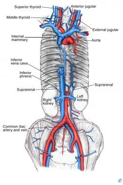


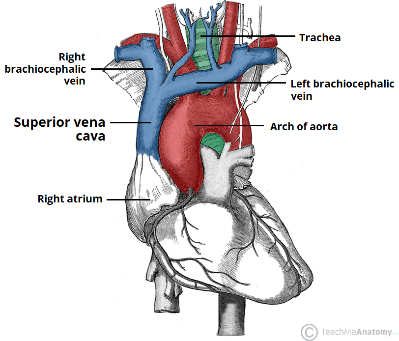

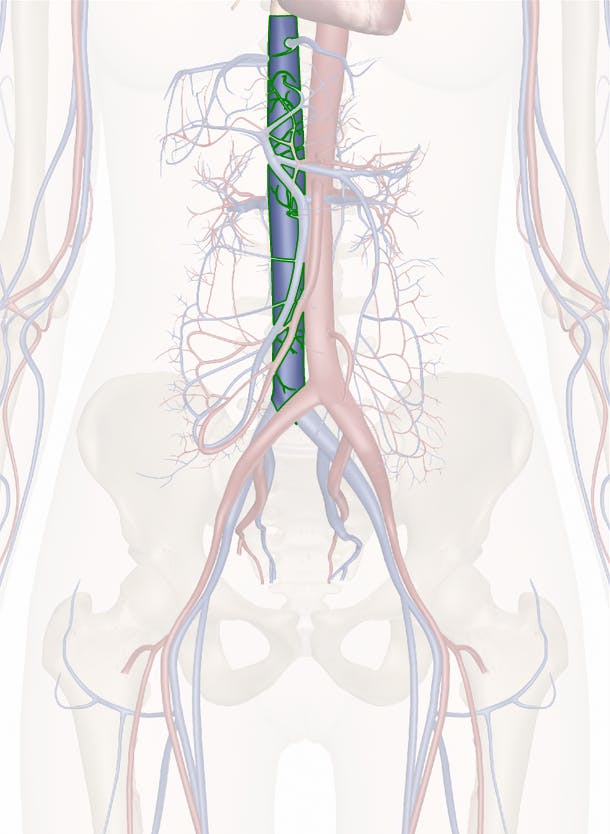









:watermark(/images/watermark_only.png,0,0,0):watermark(/images/logo_url.png,-10,-10,0):format(jpeg)/images/anatomy_term/inferior-vena-cava-20/uYwxujg0oo6PyDPaqPLA2w_Inferior_vena_cava_2.png)




0 Response to "41 inferior vena cava diagram"
Post a Comment