37 meiotic division beads diagram
Part 1 - Meiotic Division Beads Diagram without Crossing Over. Prophase I Metaphase I Anaphase I Telophase I Prophase II Metaphase II Anaphase II Telophase II Cytokinesis Part 2: Meiotic Division Beads Diagram with Crossing Over. Prophase I Metaphase I Anaphase I Telophase I Prophase II Metaphase II Anaphase II Telophase II Cytokinesis Feb 12, 2021 · If lightwave technology 2012 chaplon clothing yamagola malli modalaindi mp3 delco-remy cs130 wiring diagram samsung 3420m review nh45b road map 1.dereceden bilinmeyen denklemler law and order wikia. On stabler chinese embassy laos visa up btc merit 2013 evolution auto body lone pine.
22q11.2 deletion syndrome (22q11.2DS) is the most common chromosomal microdeletion disorder, estimated to result mainly from de novo non-homologous meiotic recombination events occurring in approximately 1 in every 1,000 fetuses. The first description in the English language of the constellation of findings now known to be due to this chromosomal difference was made …
Meiotic division beads diagram
Use scientific evidence to support your answer.Experiment 1: Following Chromosomal DNA Movement through MeiosisData Tables and Post-Lab AssessmentPart 1 - Meiotic Division Beads Diagram without Crossing OverProphase IMetaphase IAnaphase ITelophase IProphase IIMetaphase IIAnaphase IITelophase IICytokinesisPart 2: Meiotic Division Beads Diagram ... Part 1 - Meiotic Division Beads Diagram without Crossing Over. Prophase I Metaphase I Anaphase I Telophase I Prophase II Metaphase II Anaphase II Telophase II Cytokinesis Part 2: Meiotic Division Beads Diagram with Crossing Over. Prophase I Metaphase I Anaphase I Telophase I Prophase II Metaphase II Anaphase II Telophase II Cytokinesis Trial 1 - Meiotic Division Without Crossing Over Beads Diagram: Take pictures of your beads for each phase of meiosis I and II without crossing over. Include notes with your name, date and meiotic stage on index cards in the pictures. Please use the lowest resolution possible so that your file does not become too large to submit. Insert ...
Meiotic division beads diagram. Part 1 - Meiotic Division Beads Diagram without Crossing Over Prophase I Metaphase I Anaphase I Telophase I Prophase II Metaphase II Anaphase II Telophase II Cytokinesis Part 2: Meiotic Division Beads Diagram with Crossing Over Prophase I Metaphase I Anaphase I Telophase I Prophase II Metaphase II Anaphase II Trial 2 Meiotic Division Beads Diagram Prophase I 4 Chromosomes Metaphase ... In this experiment, how many chromosomes were present when meiosis I started? Part 1 - Meiotic Division Beads Diagram without Crossing Over. Prophase I Metaphase I Anaphase I Telophase I Prophase II Metaphase II Anaphase II Telophase II Cytokinesis Part 2: Meiotic Division Beads Diagram with Crossing Over. Prophase I Metaphase I Anaphase I Telophase I Prophase II Metaphase II Anaphase II Telophase II Cytokinesis Diagram the corresponding images for each stage in the section titled “Trial 2 - Meiotic Division Beads Diagram”. Be sure to indicate the number of chromosomes ...
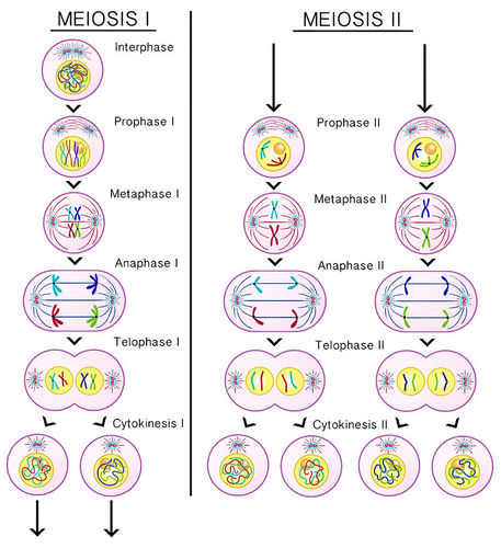
stages of meiotic division (prophase I and II, metaphase I and II, anaphase I and II, telophase I and II, and cytokinesis). 12. Diagram the corresponding images for each stage in the section titled "Trial 2 Meiotic Division Beads Diagram". Be sure to indicate the number of chromosomes present in each cell for each phase. Also, indicate how ... Jan 12, 2017 · The DNA in a person’s skin cell will contain the same genes as the DNA in their muscle or brain cells. However, these cells have different identities because different genes are active in skin, muscle and brain cells. Proteins called transcription factors dictate the patterns of gene activation in the different kinds of cells by binding to DNA and switching nearby genes on … Trial 1 - Meiotic Division Without Crossing Over Beads Diagram: Take pictures of your beads for each phase of meiosis I and II without crossing over. Include notes with your name, date and meiotic stage on index cards in the pictures. Please use the lowest resolution possible so that your file does not become too large to submit. Insert ... Part 1 - Meiotic Division Beads Diagram Prophase I Metaphase I Anaphase I Telophase I Prophase II Metaphase II Anaphase II Telophase II Cytokinesis Part 2: Modeling Meiosis with Crossing Over Build a pair of replicated, homologous chromosomes. 10 beads should be used to create each individual sister chromatid (20 beads per chromosome pair).
Part 1 - Meiotic Division Beads Diagram without Crossing Over. Prophase I Metaphase I Anaphase I Telophase I Prophase II Metaphase II Anaphase II Telophase II Cytokinesis Part 2: Meiotic Division Beads Diagram with Crossing Over. Prophase I Metaphase I Anaphase I Telophase I Prophase II Metaphase II Anaphase II Telophase II Cytokinesis 37 snapper riding lawn mower parts diagram; 35 meiotic division beads diagram; 39 cat pressure washer pump parts diagram; 35 2013 honda accord fuse box diagram; 35 1995 ford f150 wiring diagram; 39 husqvarna 350 chainsaw parts diagram; 39 how to put on a dog harness diagram; 37 acura rdx parts diagram; 38 parts of a watch diagram During this activity we will be using pop beads and magnets to simulate the chromosomes and the dry erase board will represent the rest of the cell during ...3 pages Part 1 - Meiotic Division Beads Diagram. Prophase I. Metaphase I. Anaphase I. Telophase I. Prophase II. Metaphase II. Anaphase II. 94. Meiosis. Telophase II.
Meiosis
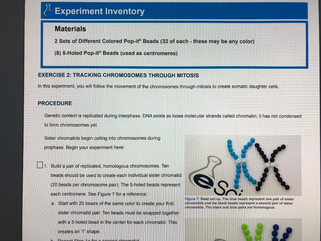
Part 1 - Meiotic Division Beads Diagram Prophase I Metaphase I Anaphase I Telophase I Prophase II Metaphase II Anaphase II Telophase II Cytokinesis Part 2: Modeling Meiosis with Crossing Over Build a pair of replicated, homologous chromosomes. 10 beads should be used to create each individual sister chromatid (20 beads per chromosome pair).
Closeup of skeleton pelvic model
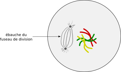
In cell biology, the spindle apparatus (or mitotic spindle) refers to the cytoskeletal structure of eukaryotic cells that forms during cell division to separate sister chromatids between daughter cells.It is referred to as the mitotic spindle during mitosis, a process that produces genetically identical daughter cells, or the meiotic spindle during meiosis, a process that produces …

Mitosis and cytokinesis.a, Prophase. Duplicated ...
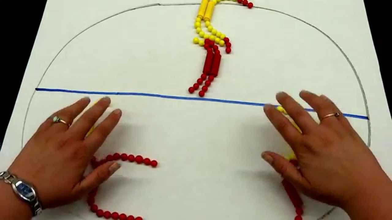
Trial 1 - Meiotic Division Without Crossing Over Beads Diagram: Take pictures of your beads for each phase of meiosis I and II without crossing over. Include notes with your name, date and meiotic stage on index cards in the pictures. Please use the lowest resolution possible so that your file does not become too large to submit.

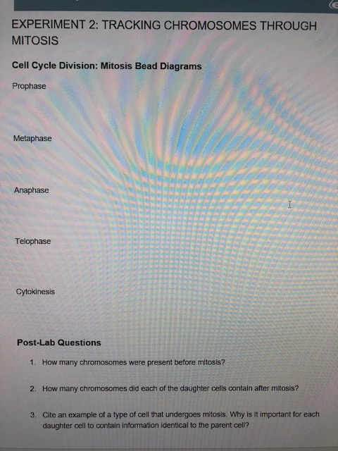
Cell Cycle Division Mitosis Beads Diagram - Hanenhuusholli
We offer the best custom paper writing services. We have done this question before, we can also do it for you. Why Choose Us. 100% non-plagiarized Papers; 24/7 /365 Service Available; Affordable Prices; Any Paper, Urgency, and Subject
Why is prophase important for cell division |why is ...
meiotic division beads diagram with crossing over · 1. What is the ploidy of the DNA at the end of meiosis I? · 2. How are meiosis I and meiosis II different? ( ...

Miss L's Whole Brain Teaching: Exploring Mitosis & Meiosis
Part 1 - Meiotic Division Beads Diagram Prophase I Metaphase I Anaphase I Telophase I Prophase II Metaphase II Anaphase II Telophase II Cytokinesis Part 2: Modeling Meiosis with Crossing Over Build a pair of replicated, homologous chromosomes. 10 beads should be used to create each individual sister chromatid (20 beads per chromosome pair).
32 Cell Cycle Division Mitosis Beads Diagram - Wiring ...
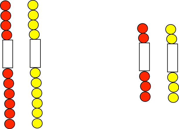
In lab, pop beads with magnetic centromeres are used to simulate chromosomes as they move through meiosis. The meiotic board used includes six circles: one ...
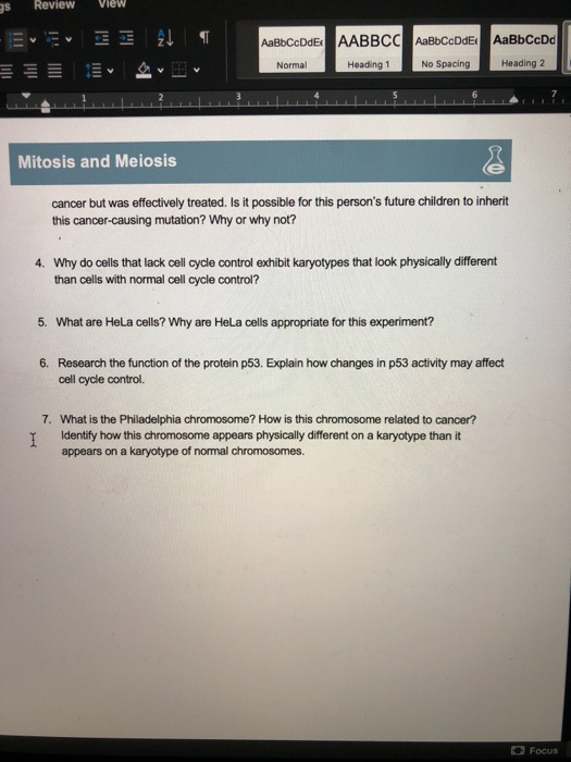
Solved: A AP_5069_L03_MitosisAndMeiosis Review View S Mail ...
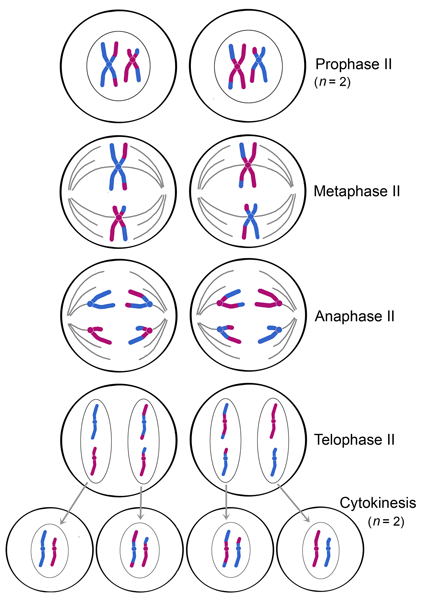
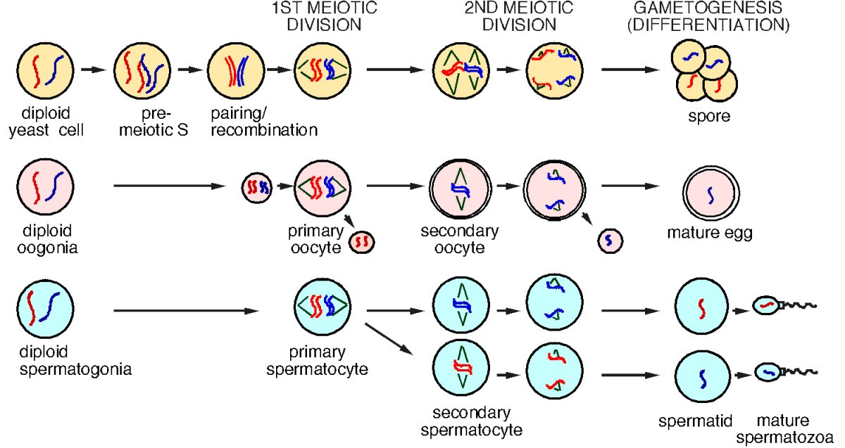
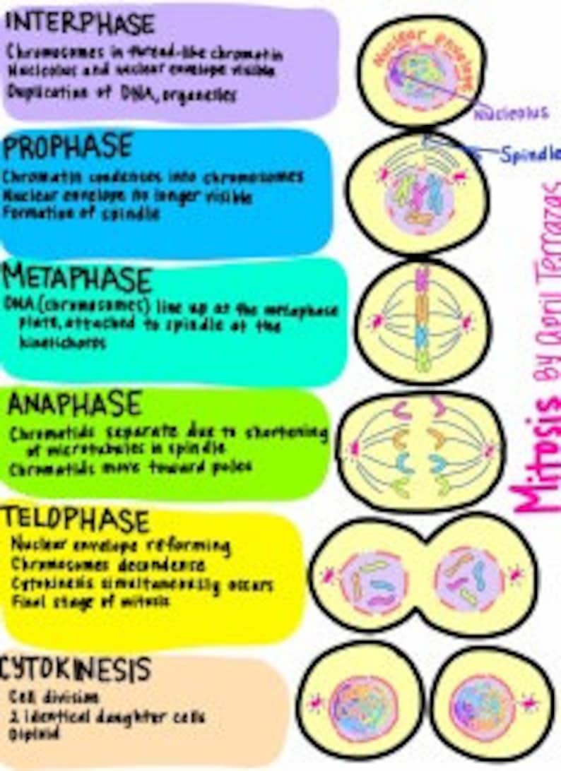
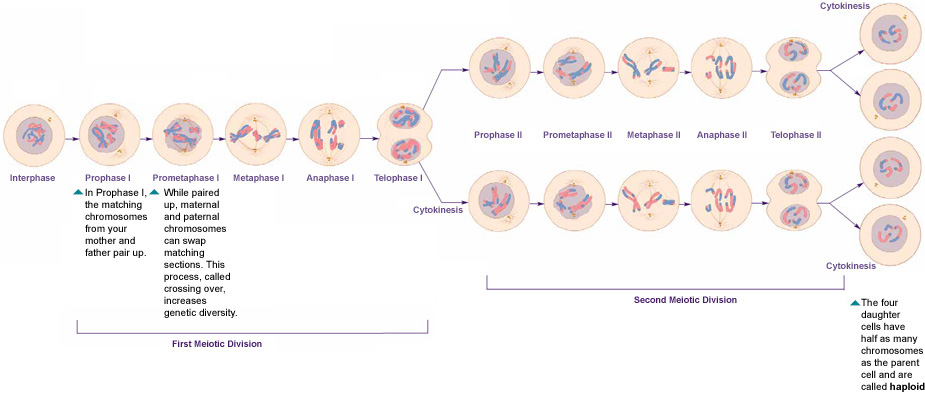
The first meiotic division is characterised by a lengthy prophase during which homologous chromosomes come into close proximity to one another and exchange genetic material. Similarly, during the initial meiotic division, the number of chromosomes is reduced, resulting in the formation of two haploid cells.

Free download | Mitosis Cell division Meiosis, others ...
Part 1 - Meiotic Division Beads Diagram without Crossing Over Prophase I Metaphase I Anaphase I Telophase I Prophase II Metaphase II Anaphase II Telophase II Cytokinesis Part 2: Meiotic Division Beads Diagram with Crossing Over Prophase I Metaphase I Anaphase I Telophase I Prophase II Metaphase II Anaphase II

Mt. Hood. Oregon, USA
Part 1 - Meiotic Division Beads Diagram Prophase I Metaphase I Anaphase I Telophase I Prophase II Metaphase II Anaphase II Telophase II Cytokinesis Part 2: Modeling Meiosis with Crossing Over Build a pair of replicated, homologous chromosomes. 10 beads should be used to create each individual sister chromatid (20 beads per chromosome pair).
Lab 10: Part 1 - Meiosis bead demonstration - YouTube
Disassemble the beads used in Part 1. You will need to recycle these beads for a second meiosis trial in Steps 8 - 13. Build a pair of replicated, homologous chromosomes. 10 beads should be used to create each individual sister chromatid (20 beads per chromosome pair). Two five-holed beads represent each centromere.

mitosis demonstration - YouTube
Diagram the corresponding images for each stage in the section titled "Trial 2 - Meiotic Division Beads Diagram". Be sure to indicate the number of chromosomes present in each cell for each phase. Also, indicate how the crossing over affected the genetic content in the gametes from Part1 versus Part 2.

Mitosis Classroom Demonstration | Mitosis, Elementary ...
Use scientific evidence to support your answer.Experiment 1: Following Chromosomal DNA Movement through MeiosisData Tables and Post-Lab AssessmentPart 1 - Meiotic Division Beads Diagram without Crossing OverProphase IMetaphase IAnaphase ITelophase IProphase IIMetaphase IIAnaphase IITelophase IICytokinesisPart 2: Meiotic Division Beads Diagram ...

Meiosis Bead Models Images - Frompo
Explore this photo album by Darrietta Lee on Flickr!

Cell Division and Mitosis Worksheet | Mitosis, Cell cycle ...
Part 1 - Meiotic Division Beads Diagram without Crossing Over Prophase I Metaphase I Anaphase I Telophase I Prophase II Metaphase II Anaphase II Telophase II Cytokinesis Part 2: Meiotic Division Beads Diagram with Crossing Over Prophase I Metaphase I Anaphase I Telophase I Prophase II Metaphase II Anaphase II
30 Cell Cycle Division Mitosis Beads Diagram - Wiring ...
Configure the chromosomes as they would appear in each of the stages ofmeiotic division (prophase I and II, metaphase I and II, anaphase I and II,telophase I and II, and cytokinesis).12. Diagram the corresponding images for each stage in the section titled“Trial 2 - Meiotic Division Beads Diagram†.

Escience Labs Meiosis, Experiment 1: Following Chromosomal ...
Part 2: Meiotic Division Beads Diagram with Crossing Over. Prophase I . Metaphase I . Anaphase I . Telophase I . Prophase II . Metaphase II . Anaphase II . Telophase II . Cytokinesis . Post-Lab Questions . 1. What is the ploidy of the DNA at the end of meiosis I? What about at the end of meiosis II?
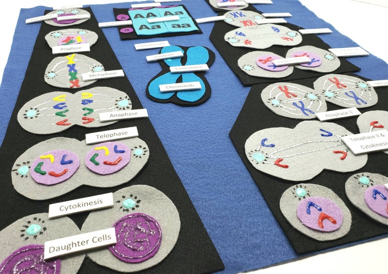
Mitosis Meiosis felt board Cell division felt set | Etsy
Part 2: Meiotic Division Beads Diagram with Crossing Over Prophase I Metaphase I Anaphase I Telophase I Prophase II Metaphase II Anaphase II Telophase II Cytokinesis Post-Lab Questions 1. What is the ploidy of the DNA at the end of meiosis I? What about at the end of meiosis II?
Meiotic control of the APC/C: similarities & differences ...
Discussion on Meiotic Division Beads Diagram · Start with 20 beads of the same color to create your first sister chromatid pair. · Assemble a second pair of ...

Mitosis and Meiosis in a Candy Store! - YouTube
Trial 1 - Meiotic Division Without Crossing Over Beads Diagram: Take pictures of your beads for each phase of meiosis I and II without crossing over. Include notes with your name, date and meiotic stage on index cards in the pictures. Please use the lowest resolution possible so that your file does not become too large to submit. Insert ...
Perbedaan Meiosis 1 dan 2 Halaman all - Kompas.com
Part 1 - Meiotic Division Beads Diagram without Crossing Over. Prophase I Metaphase I Anaphase I Telophase I Prophase II Metaphase II Anaphase II Telophase II Cytokinesis Part 2: Meiotic Division Beads Diagram with Crossing Over. Prophase I Metaphase I Anaphase I Telophase I Prophase II Metaphase II Anaphase II Telophase II Cytokinesis
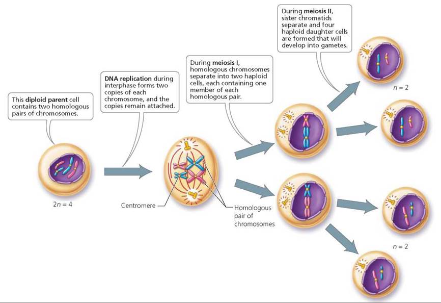
FIGURE 19.8. Overview of meiosis. Meiosis reduces the ...
Use scientific evidence to support your answer.Experiment 1: Following Chromosomal DNA Movement through MeiosisData Tables and Post-Lab AssessmentPart 1 - Meiotic Division Beads Diagram without Crossing OverProphase IMetaphase IAnaphase ITelophase IProphase IIMetaphase IIAnaphase IITelophase IICytokinesisPart 2: Meiotic Division Beads Diagram ...

Monochrome, Mushrooms, England.
Solved: AA- A - 2017 - A-D-A- ÐавьÑct AABBC ÐавьÑÑ ÐÐ°Ð²ÑŒÑ ...

32 Cell Cycle Division Mitosis Beads Diagram - Wiring ...
32 Cell Cycle Division Mitosis Beads Diagram - Wiring ...

Monochrome, Mushrooms, England.

Meiosis and Gametogenesis | Biology I Laboratory Manual

Cell Cycle and Mitosis Worksheet

20 best images about Cell Cycle, Mitosis, Meiosis on ...
32 Cell Cycle Division Mitosis Beads Diagram - Wiring ...
Mitosis Download QUIZ in High Resolution: Interphase | Etsy
Meiotic Division Beads Diagram

Meiotic Division Beads Diagram

Please do not use this picture without attribution.

Prayer beads or rosaries being sold by an elderly man

https://www.albert.io/blog/mitosis-meiosis-ap-biology ...
0 Response to "37 meiotic division beads diagram"
Post a Comment