38 spine l5 s1 diagram
10/08/2011 · Roussouly P, Gollogly S, Berthonnaud E, et al. Sagittal alignment of the spine and pelvis in the presence of L5-S1 isthmic lysis and low-grade spondylolisthesis. Spine. 2006; 31:2484–2490. doi: 10.1097/01.brs.0000239155.37261.69. [Google Scholar] The lumbo-sacral spine includes: Lumbar vertebrae: Numbered L1 through L5, these odd-shaped vertebrae signal the end of the typical bones of the spinal column.; Sacrum: This triangle-shaped bone ...
http://www.johngibbonsbodymaster.co.ukJohn Gibbons is a registered Osteopath, Lecturer and Author and is demonstrating how to manipulate (HVT - Grade 5) the ...

Spine l5 s1 diagram
The five vertebrae of the lumbar spine are connected in the back by facet joints, which allow for forward and backward extension, as well as twisting movements. The two lowest segments in the lumbar spine, L5-S1 and L4-L5, carry the most weight and have the most movement, making the area prone to injury. Lumbar Spine. The lumbar spine has 5 vertebrae, which are numbered from L1 through L5. The lumbar vertebrae are the largest bones in the spinal column and support a great deal of body weight. Sacrum. The sacrum has five bones, which are fused together in adults and are numbered S1 through S5. It connects the spine to the hipbones. Coccyx OSMOSIS.ORG 709. This Osmosis High-Yield Note provides an overview of Spinal disorders essentials. All Osmosis Notes are clearly laid-out and contain striking images, tables, and diagrams to help visual learners understand complex topics quickly and efficiently. Find more information about Spinal disorders by visiting the associated Learn Page.
Spine l5 s1 diagram. The sacrum is the triangle-shaped bone at the end of the spine between the lumbar spine and the tailbone. The sacral spine consists of five segments, S1 - S5, that together affect nerve communication to the lower portion of the body. It is important to understand that the spinal cord does not extend beyond the lumbar spine. Official Ninja Nerd Website: https://ninjanerd.orgNinja Nerds!In this lecture Professor Zach Murphy will present on the lumbar vertebrae (L1-L5) anatomy thro... The fifth lumbar spine vertebrae (L5) is part of the greater lumbar region. To the human eye, this is the curve just above the buttocks, which is also commonly referred to as the small of the back ... The lumbar spine (lower back) consists of five vertebrae in the lower part of the spine, between the ribs and the pelvis. Lumbar spinal stenosis is a narrowing of the spinal canal, compressing the nerves traveling through the lower back into the legs. While it may affect younger patients, due to developmental causes, it is more often a degenerative condition that affects people who are ...
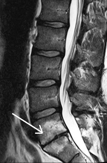
19/05/2021 · Furthermore, we attached it along the spine, including S1–L1, L1/T12–C7, C1–C7, and S1–C7 (C, T, L, and S represent the cervical, thoracic, lumbar, and sacrum segments of … The L5 nerve to the top of the foot and big toe. The S1 nerve to the outside and bottom of the foot. The disc normally is composed of 2 parts. These are microscopic and cannot exactly be differentiated on an MRI. The central softer part of the disc is the nucleus and the out layer is the annulus. Download scientific diagram | Pre-operative MRI lumbar spine showing a L5/S1 prolapse. from publication: Positioning a proned patient with cauda equina syndrome who presents at 15 weeks gestation ... Spinal Cord Diagram at the Canadian Paraplegic Association (NS) web site. Spinal Cord Anatomy Apparelyzed.com. This series of eight guides describes outcomes according to level of spinal cord injury (C1-3, C4, C5, C6, C7-8, T1-9, T10-L1 and L2-S5). Each guide provides individual guidance on what people with different levels of SCI can ...
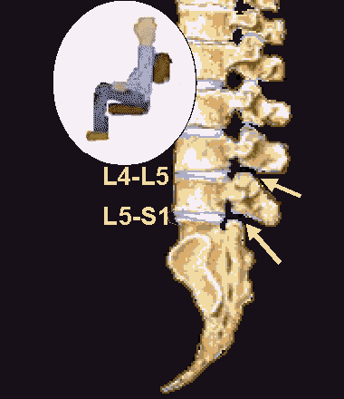
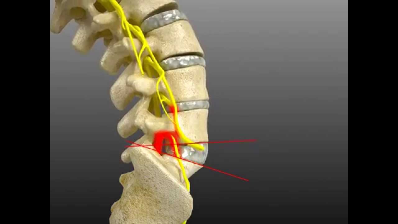
The twelve thoracic vertebrae are numbered T1 to T12. The range of motion in the thoracic spine is limited. Lumbar (low back) - the main function of the lumbar spine is to bear the weight of the body. The five lumbar vertebrae are numbered L1 to L5. These vertebrae are much larger in size to absorb the stress of lifting and carrying heavy objects. L5-S1 is the exact spot where the lumbar spine ends and the sacral spine begins. The lumbosacral joint is the joint that connects these bones. L5-S1 is composed of the last bone in the low back, called L5, and the triangle-shaped bone beneath, known as the sacrum. The sacrum is made of five fused bones, of which the S1 is the topmost. Anatomy The fifth lumbar spinal nerve 5 (L5) originates from the spinal column from below the lumbar vertebra 5 (L5). L5 supplies many muscles, either directly or through nerves originating from L5. They are not innervated with L5 as single origin, but partly by L5 and partly by other spinal nerves. The muscles are: gluteus maximus muscle mainly S1 The lumbosacral joint (L5-S1) is a joint in the spinal column between the last vertebra of the lumbar region and the first vertebra of the sacral region. At this point, there is a shift in the curvature of the vertebral column from a forward curvature in the lumbar region to a backward curvature in the sacral section of the spinal column.
A-MRI lumbosacral spine saggital section showing extruded ...
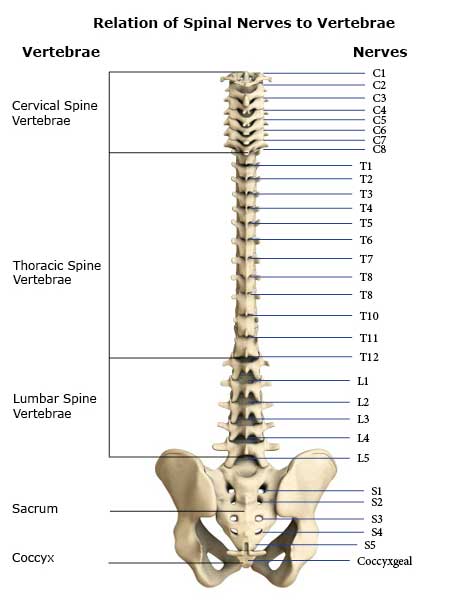
Spine Diagrams. The human spine consists of 33 vertebrae: 7 Cervical vertebra (C1-C7) 12 Thoracic vertebra (T1-T12) 5 Lumbar vertebra (L1-L5) The sacrum and coccyx are made up of 9 fused vertebrae. Each vertebra is attached to the one above and below it by ligaments and muscles.

Diagram of Lifting | Physics of Lifting | Common Spine Injuries | Denver, Colorado Spine Doctor
Anatomical diagrams of the spine and back. This human anatomy module is composed of diagrams, illustrations and 3D views of the back, cervical, thoracic and lumbar spinal areas as well as the various vertebrae. It contains the osteology, arthrology and myology of the spine and back. It is particularly interesting for physiotherapists ...

Image 10129: Discectomies and Fusion at L5-S1 Illustration ...
The L5-S1 spinal motion segment, also called the lumbosacral joint, is the transition region between the lumbar spine and sacral spine in the lower back. In this region, the curvature of the spine changes from lumbar lordosis (forward curve) to sacral kyphosis (backward curve). L5-S1 helps transfer loads from the spine into the pelvis and legs.

Spinal cord injury C3/4, C4/5, C5/6 T12 L3, L4, L5, S1 ...
The lumbar spine makes up the the lower end of the spinal column. It consists of 5 lumbar vertebra that are numbered 1 through 5 from top to bottom i.e. L1, L2, L3, L4, and L5. The L5 vertebra is connected to the top of the sacrum (named the S1 segment) through an intervertebral disc. To review, the purpose of the spine, as a whole, is to ...

Discogram of Lumbar Spine at L3-4, L4-5 & L5-S1
Spinal stenosis occurs most often in the lower back and the neck. Some people with spinal stenosis may not have symptoms. Others may experience pain, tingling, numbness and muscle weakness. Symptoms can worsen over time. Spinal stenosis is most commonly caused by wear-and-tear changes in the spine related to osteoarthritis.

Body Aches because corona symptom
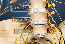
Lower back, anatomically lumbo-sacral spine is the most vulnerable part of the spine. Because of the nature of the movement as well as structure and shape of the spine. There are certain causes behind problems of L3-L4, L4-L5 and L5-S1 discs. Muscular Stiffness and disc problems in lower back

Qu'est ce qu'une sciatique ? | Santé Orthopédique
The L5 vertebrae sits on top of the S1 vertebrae. Compression of the nerve root between L5 and S1 vertebrae can cause pain, numbness, tingling and weakness in the leg on the affected side. Core strengthening can help relieve the symptoms of L5-S1 compression. exercises to relieve l5 s1 compression.
Patient with a disc herniation (white arrow) at level L5 ...
Low Back Pain - L4 5 and L5 S1 Disc Bulges Progressing to Disc Herniations with Spinal Fusion Surgery L5-S1 Disc Herniation and Shoulder Injury with Subsequent Repair of Shoulder. L5-S1 Disc Herniation Progression - Superior View. L5-S1 Laminectomy, Partial ...
![[DIAGRAM] Diagram Of L4 L5 FULL Version HD Quality L4 L5 ...](http://c7.alamy.com/comp/ADW9JC/low-back-pain-l4-5-and-l5-s1-disc-bulges-progressing-to-disc-herniations-ADW9JC.jpg)
[DIAGRAM] Diagram Of L4 L5 FULL Version HD Quality L4 L5 ...
the diagram at the right. Multiple sclerosis (MS) is a potentially disabling disease of the brain and spinal cord (central nervous system). In MS, the immune system attacks the protective sheath (myelin) that covers nerve fibers and causes communication problems between your brain and the rest of your body.
Consequences of Office Chair Sitting (2): The Kink at the ...
Pain. Pain is a common symptom associated with L5-S1 pinched nerves 3.This may feel like a dull ache or a sharp pain. L5 nerve compression causes pain along the outer border of the back of your thigh, while S1 nerve compression causes pain in your calf and the bottom of your foot 3. This pain can range from mild to severe and may be constant or intermittent.

a Case 7: male, 52 years old. Preoperative MRI: severe DDD ...
Spondylolisthesis: Diagram of L5 vertebra sitting correctly on the sacrum. Written by Mary Rodts, DNP. fig. 1. Diagram of an L5 vertebra "sitting" corrtectly on the sacrum. fig. 1a. Diagram of an L5 vertebra slipping forward on the sacrum (i.e., spondylolisthesis) Updated on: 02/01/10.

MRI showing L4-L5 and L5-S1 degenerative disc disease ...
Back pain or leg pain can typically arise due to injury where the lumbar spine and sacral region connect (at L5 - S1) because this section of the spine is subjected to a large amount of stress and twisting. People with rheumatoid arthritis or osteoporosis are inclined to develop stress fractures and fatigue fractures in the sacrum.

Radiographic progress of L5-S1 fusion at 18 months ...
This is the case of a 28-year-old male who suffered from urinary frequency, perineal pain and mild low back and buttock pain. An MRI confirmed a lateral L5 disc bulge and a fixation at L5/S1. After two adjustments to the 5th lumbar vertebrae the patient’s pelvic …

The spinal mass localized at L4-L5-S1. | Download ...
The sacral vertebrae—also called the sacral spine—consists of five sacral vertebrae bones. These bones fuse together to form the sacrum, the shield-shaped bony structure located at the base of the lumbar vertebrae (the five cylindrical bones forming the spine of the lower bank) and connected to the pelvis. The sacral vertebrae are represented by segments S1 through S5 and located between ...
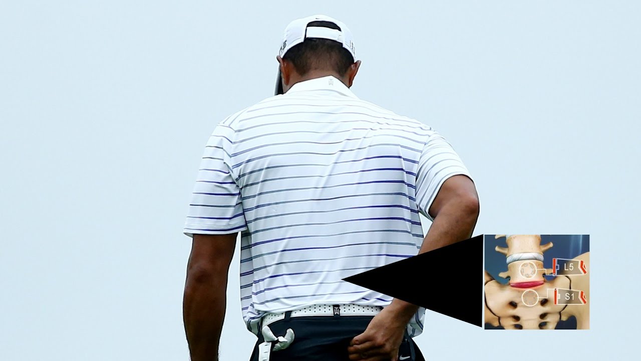
A look at Tiger Woods' L5/S1 spinal fusion back surgery ...
Lumbar vertebrae. Position of human lumbar vertebrae (shown in red). It consists of 5 bones, from the top down, L1, L2, L3, L4 and L5. The lumbar vertebrae are, in human anatomy, the five vertebrae between the rib cage and the pelvis. They are the largest segments of the vertebral column and are characterized by the absence of the foramen ...

MRI scan showing massive L5/S1 disc protrusion. | Download ...
07/09/2021 · Neurological manifestations are reported in up to 36% of patients with COVID-19. Among COVID-19-associated CNS conditions, GBS has emerged in an increasing number of case reports as an additional hazard with a significant risk of mortality or prolonged respiratory failure.4,11,12,14–20 We herein present an in-depth systematic review of COVID-19–related …

CT scan sagittal views showing the right L5/S1 foraminal ...
The lumbar spine is the lower back that begins below the last thoracic vertebra (T12) and ends at the top of the sacral spine, or sacrum (S1). Most people have 5 lumbar levels (L1-L5), although it is not unusual to have 6. Each lumbar spinal level is numbered from top to bottom—L1 through L5, or L6.

Parasagital computed tomography slice. Note: L5-S1 ...
OSMOSIS.ORG 709. This Osmosis High-Yield Note provides an overview of Spinal disorders essentials. All Osmosis Notes are clearly laid-out and contain striking images, tables, and diagrams to help visual learners understand complex topics quickly and efficiently. Find more information about Spinal disorders by visiting the associated Learn Page.

Pin em Anatomy
Lumbar Spine. The lumbar spine has 5 vertebrae, which are numbered from L1 through L5. The lumbar vertebrae are the largest bones in the spinal column and support a great deal of body weight. Sacrum. The sacrum has five bones, which are fused together in adults and are numbered S1 through S5. It connects the spine to the hipbones. Coccyx

(A) Sagittal T2 MRI demonstrating L5-S1 disc extrusion. (B ...
The five vertebrae of the lumbar spine are connected in the back by facet joints, which allow for forward and backward extension, as well as twisting movements. The two lowest segments in the lumbar spine, L5-S1 and L4-L5, carry the most weight and have the most movement, making the area prone to injury.
Trapped nerves in the Lower Back

#spine | Spinal nerve, Spine health, Nerves function

What Is L5 S1 Degenerative Disc Disease - Captions Imajinative
All about L5-S1 (Lumbosacral Joint)

Low Back Pain - Disc Herniation ,Sciatica - Everything You ...
Causes of Lower Back Pain | Orthopedic Spine Surgeon | CO

Spinal Cord Injury Levels | Bone and Spine | Physical ...
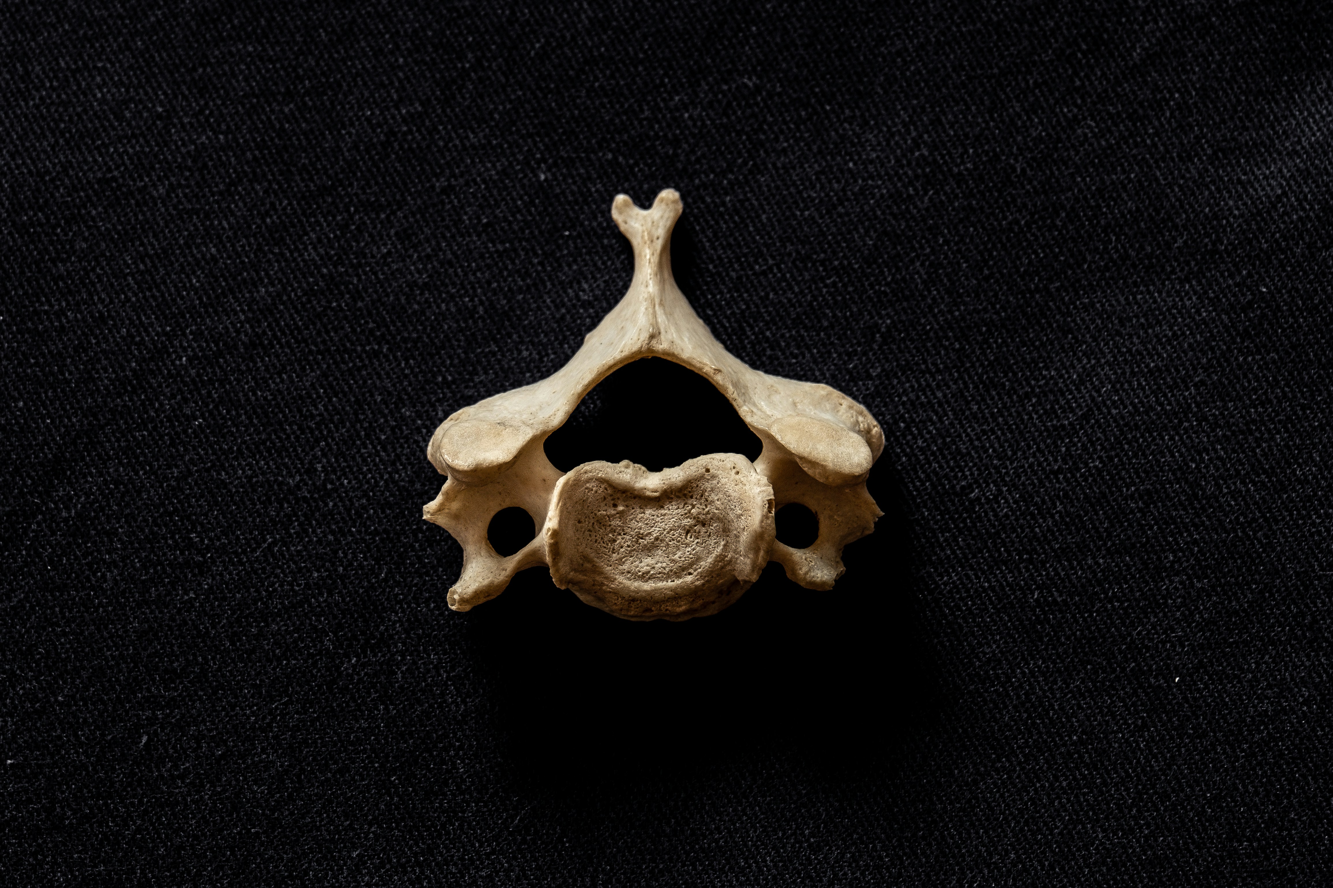
Typical C3-6, human cervical vertebra , bifid spinous process clearly visible

celinasews: Spinal Nerve Chart Fun | Physical therapy ...

Spinal Fusion Surgery - L5-S1 Spondylolisthesis with ...

T1 (a) and T2 (b) sagittal lumbar MRI showing an L5/S1 ...

Anteroposterior (A) and lateral (B) standard X-rays of L4 ...

Problems with the L5-S1 Disc?
Spinal Adjustment Treatment for L4/L5 Fixation Causing ...

L5 S1 Anatomy

Colorful cactus 🌵

Aspect of pseudotumoral L5-S1 Charcot spine in a C5 ...

Pin by sara dernvall on Spondylolisthesis Awareness By ...
0 Response to "38 spine l5 s1 diagram"
Post a Comment