37 sheep brain diagram labeled
12 Best Images of Brain Anatomy Worksheet - Brain Diagram ... By the way, concerning Brain Anatomy Worksheet, below we can see particular variation of photos to add more info. brain diagram worksheet, brain diagram and functions worksheet and labeled sheep brain worksheet are three main things we will show you based on the gallery title. A virtual sheep brain dissection guides anatomy studies ... Jun 6, 2018 - A virtual sheep brain dissection guides anatomy studies with photos & blank diagrams. Also shop complete dissection kits: guide, tools & preserved specimen.
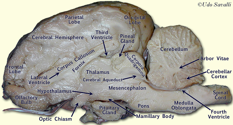
Sheep Brain Dissection with Labeled Images The sheep brain is exposed and each of the structures are labeled and described in a sequential manner, in the same way that a real dissection would occur. Sheep Brain Dissection with Labeled Images Sheep Brain Dissection 1. The sheep brain is enclosed in a tough outer covering called the dura mater.

Sheep brain diagram labeled
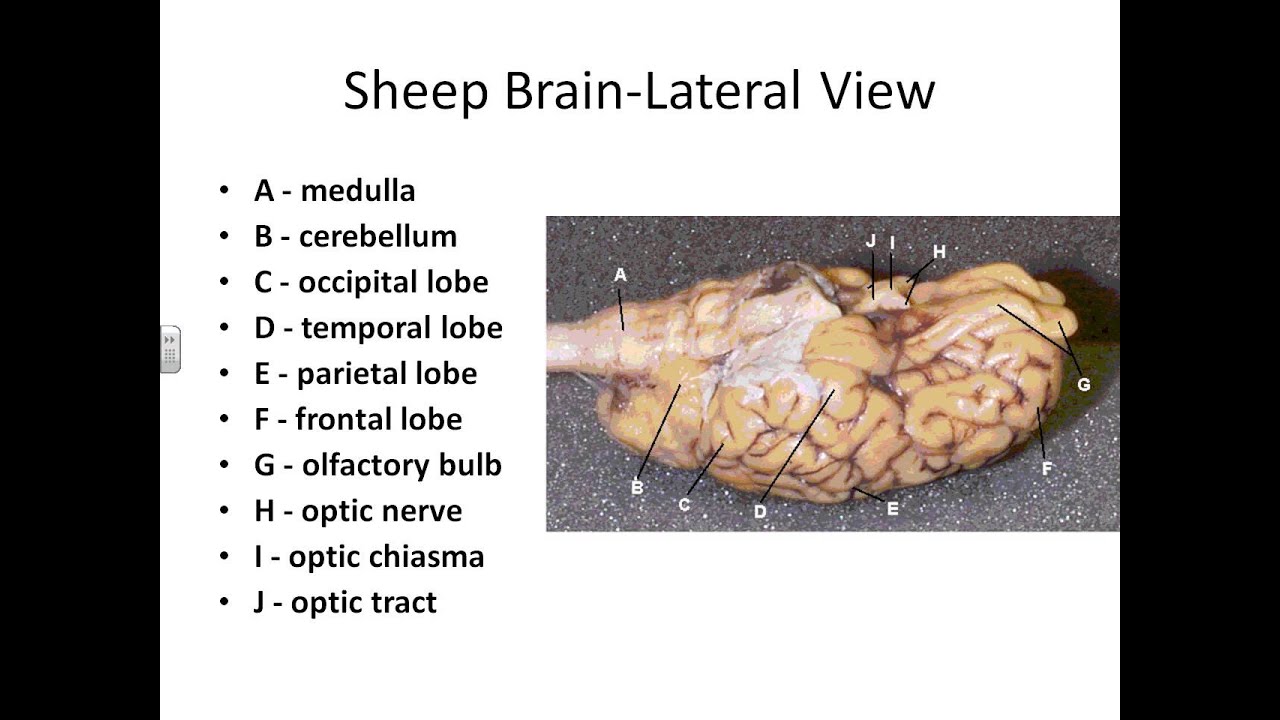
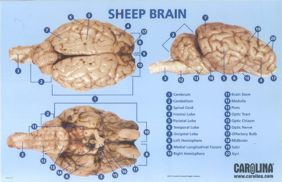
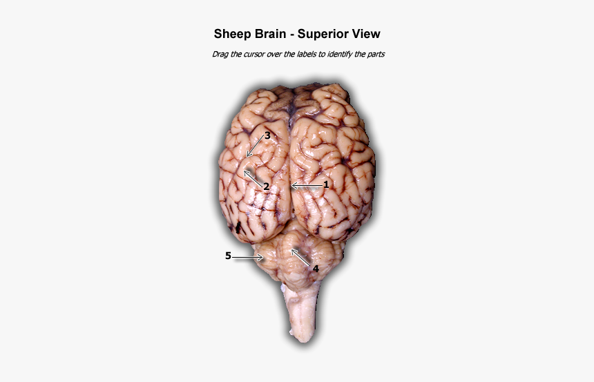
PDF Sheep Neuroanatomy Lab- Labeling Worksheet Psychology 2315 ... Sheep Neuroanatomy Lab- Labeling Worksheet Psychology 2315- Brain and Behaviour Kwantlen Polytechnic University Figure 1: Dorsal view Cerebellum, Frontal lobe, Occipital lobe, Parietal lobe, and Temporal lobe. Temporal Parietal Lobe Frontal Lobe Cerebellum Occipital Lobe Functions - Sheep Brain Dissection Hypothalamus- is a portion of the brain that contains a number of small nuclei with a variety of functions. One of the most important functions of the hypothalamus is to link the nervous system to the endocrine system via the pituitary gland. Cerebral Acquesduct-a fluid-filled canal that runs through the midbrain connecting the third and fourth ... Sheep Brain Neuroanatomy Online Self-Test | KPU.ca ... Sheep Brain Neuroanatomy Online Self-Test. Use each diagram as a reference, and selected the correct answer for each lettered structure. You may find it useful to open the diagrams in a separate window to review while answering each question. Dorsal Surface.
Sheep brain diagram labeled. PDF L2 StudentPacket SheepBrainExploration Why$dread$a$bump$on$the$head?$ $ October$2012$ Lesson$2:$What$does$thebrain$looklike?$ $ $ $ 4 $ PlanesofOrientation ... Sheep Brain Diagram - Diagram Sketch Sheep Brain Diagram. angelo on October 9, 2021. Image Result For Sheep Brain Labeled Brain Diagram Human Brain Diagram Brain Anatomy. Sheep Brain Dissection Guide With Pictures Worksheets Nervous System Anatomy Brain Anatomy Dissection. Sheep Brain External View Labeled Anatomia Veterinaria Anatomia Veterinaria. › 46928068 › Why_Zebras_Dont_GetWhy Zebras Don't Get Ulcers. Robert Sapolsky - Academia.edu There has been a revolution in medicine concerning how we think about the diseases that now afflict us. It involves recognizing the interactions between the body and the mind, the ways in which emotions and personality can have a tremendous impact on PDF SHEEP BRAIN LAB Purpose: References: Instructions ... 1. Obtain a sheep brain dissection chart from your teacher. 2. Working in a group of two or three, select a whole sheep brain. Lay the brain on the dissecting tray to view the external surface. 3. Identify the external structures indicated below on the brain. Then label the external anatomy diagram with all the structures.
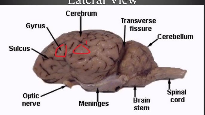
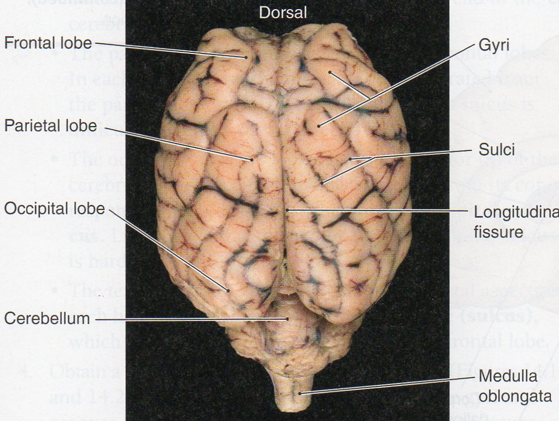
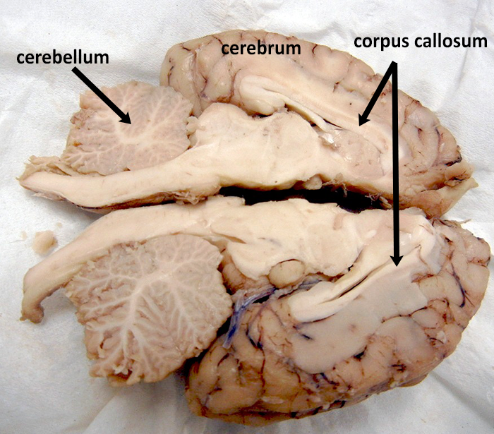
Sheep Brain Dissection - scienceteacherprogram.org o Label sheep brain diagram. o Match brain region to its function. o Application questions - could answered orally, as a written summative assignment or expanded into a research project § Show images of different mammalian brains and ask students to · 1. Label the different regions/lobes · 2. Hypothesize about the animal's capabilities ... 37 labeled sheep brain diagram - Diagram Online Source Start studying Sheep Brain Dissection labeled.Learn vocabulary, terms, and more with flashcards, games, and other study tools. function, and pathology. Those students participating in Sheep Brain Dissections will have the opportunity to dissect and compare anatomical structures. At the end of this document, you will find anatomical diagram s, vocabulary review, and pre/post tests for your ... Sheep Brain Dissection Project Guide | HST Learning Center Use the labeled picture to identify the corpus callosum, medulla, pons, midbrain, and the place where the pituitary gland attaches to the brain. (In many preserved specimens the pituitary gland is no longer present. It is not pictured.) Use your fingers or a teasing needle to gently probe the parts and see how they are connected to each other. en.wikipedia.org › wiki › MammalMammal - Wikipedia Mammals (from Latin mamma, 'breast') are a group of vertebrates constituting the class Mammalia (/ m ə ˈ m eɪ l i ə /), characterized by the presence of mammary glands which in females produce milk for feeding (nursing) their young, a neocortex (a region of the brain), fur or hair, and three middle ear bones.
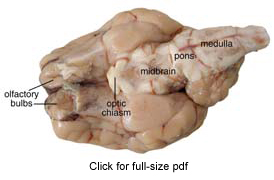
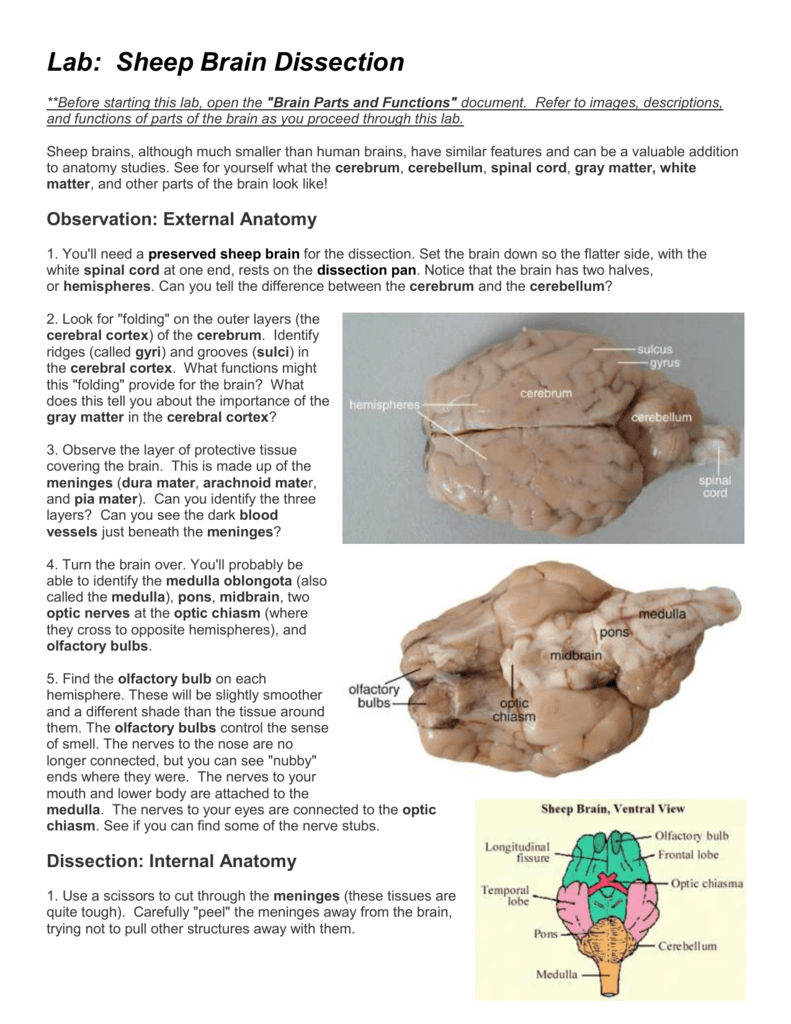
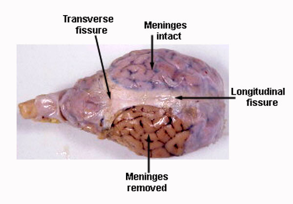
labeled brain | Brain anatomy, Anatomy and physiology, Anatomy Sheep Brain Dissection Guide. In this lab guide, students are given instruction on how to remove the dura mater, and locate the main structures of the external and internal brain. bekahaguiar. B. Becky Aguiar. School-Science. Human Brain Anatomy. Human Anatomy And Physiology. Brain Science. PDF Lab: Sheep Brain Dissection - Mrs. Moretz's Science Site Slice through the brain along the center line, starting at the . cerebrum. and going down through the . cerebellum, spinal cord, medulla, and . pons. Separate the two halves (hemispheres) of the brain and lay them with the inside facing up. 3. Use the labeled pictureand information below to identify the parts/regions of the brain as well as the . hypothalamus study.com › learn › lessonFrog Anatomy Diagram & Structure | Parts of a Frog - Video ... Jan 27, 2022 · Frog Anatomy. It is a common practice in high school biology classes to dissect a frog. Dissection is a great way to explore the anatomy of an animal. PDF DISSECTION OF THE SHEEP'S BRAIN - Hanover College Sheep Brain Dissection Guide 3. Examine the ventral surface of the sheep brain. The next several steps will view this surface of the brain. A pair of olfactory bulbs may be seen, one under each lobe of the frontal cortex. Several important parts of the visual system are visible in the ventral view of the brain. Muscles, other nerves and fatty tissue may

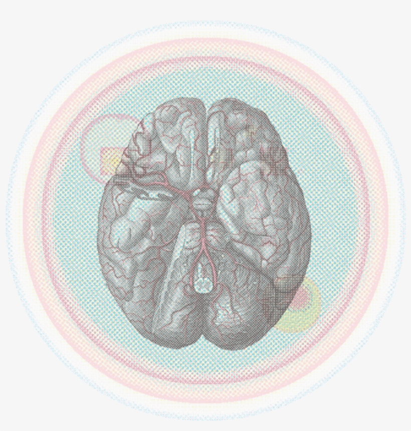
Sheep Brain Images
Sheep Brain Dissection | Human Anatomy Quiz - Quizizz Play this game to review Human Anatomy. Name this part of the brain. Preview this quiz on Quizizz. Name this part of the brain. Sheep Brain Dissection DRAFT. 6th - 12th grade. 193 times. Biology, Other Sciences. 77% average accuracy. a year ago. mrsturmscience. 0. Save. Edit. Edit. Sheep Brain Dissection DRAFT. a year ago. by mrsturmscience ...

Pretty good picture of the sheep brain labeled. | Basic ...
Sheep Brain Dissection labeled Diagram | Quizlet Start studying Sheep Brain Dissection labeled. Learn vocabulary, terms, and more with flashcards, games, and other study tools.
Sheep Brain Dissection
Cranial Nerves of the Sheep Brain Diagram | Quizlet Cranial Nerves of the Sheep Brain Diagram | Quizlet. Start studying Cranial Nerves of the Sheep Brain. Learn vocabulary, terms, and more with flashcards, games, and other study tools. Search.

Cerebrum Sheep Dissection - Human Anatomy - GUWS Medical
en.wikipedia.org › wiki › RobotRobot - Wikipedia The word robot can refer to both physical robots and virtual software agents, but the latter are usually referred to as bots. There is no consensus on which machines qualify as robots but there is general agreement among experts, and the public, that robots tend to possess some or all of the following abilities and functions: accept electronic programming, process data or physical perceptions ...
Sheep Brain Dissection Project Guide | HST Learning Center
Sheep Brain - Dorsal View Brain Anatomy IntroductionCLOSE Dissected Sheep Brain — Dorsal View Dorsal view of sheep brain with the cerebellum and caudal cerebrum removed. The rostral colliculus(large arrow label) and the caudal colliculus(small arrow label) together form the tectumof the midbrain.
The Brain - SCIENTIST CINDY
Brain Lobes Diagram Labeled - Studying Diagrams Each hemisphere has a frontal temporal parietal and occipital. With more related things such sheep brain diagram labeled brain nervous system worksheet and blank heart diagram. The diagram of the brain is useful for both Class 10 and 12. THE LOBES Occipital lobe Lower back of the brain.
BRAIN ANATOMY: 101 SHEEP BRAIN DISSECTION!
Sheep brain labeling Quiz - PurposeGames.com This online quiz is called Sheep brain labeling. This game is part of a tournament. You need to be a group member to play the tournament

sheep brain | Annette's Vet Student Info
Labeled Diagrams of the Human Brain You'll Want to Copy ... Let us have a look at these three layers and learn about the human brain diagram and functions. Human Brain: Diagram and Functions. The human brain is an astonishing organ that takes care of each function and action of the body. All the functions are carried out without a single glitch and before you even bat an eyelid. The following are the different regions of the human brain and their functions. Labeled Diagrams of the Human Brain
Sheep Brain Dissection Guide - YouTube
Sheep Brain Quiz - PurposeGames.com This is an online quiz called Sheep Brain. This quiz has tags. Click on the tags below to find other quizzes on the same subject.
Lab - Sheep Brain Dissection (word)
Sheep Brain Dissection Labeled Diagram - Studying Diagrams Sheep Brain Dissection Labeled Diagram. Place this on your dissection tray. Place the brain with the curved top side of the cerebrum facing up. By the way concerning Brain Anatomy Worksheet below we can see particular variation of photos to add more info. Identify important parts of the sheep brain in a preserved specimen.

sheep brain labeling #2 Diagram | Quizlet
35+ Sheep Brain Diagram Labeled Images - Cara Menghasilkan ... Use each diagram as a reference, and selected the correct answer for each lettered structure. Week 1 week 2 week 3 week 4. Test your knowledge on this science quiz to see how you do and. Sheep brain dissection is a mainstay of many neuroscience and biological psychology lab courses. Anatomy diagrams blank brain diagram inspirational blank brain ...
Sheep Brain Dissection Picture Guide
Sheep Brain Labeled Diagram - Diagram Sketch Labeled Sheep Brain Diagrams Hol Brain Anatomy Nervous System Anatomy Basic Anatomy And Physiology. Sheep Brain Dissection Project Guide Hst Learning Center Dissection Life Science Lessons Brain Based Learning. Sheep Brain Dissection Lab Companion In 2021 Brain Anatomy Anatomy And Physiology Brain.
Sheep+brain+dissection
PDF Neuroanatomy: Dissection of the Sheep Brain brain from the pan and place it dorsal side down on some paper towels. You are going to bisect the brain along the orientation of the longitudinal fissure. Align the brain so that it is not angled and you will achieve a symmetrical cut. Bisect the frontal lobes, optic chiasm, the mammillary bodies, the pons and the medulla. Try to cut with one smooth
Online A&P I Lab Sheep Brain Dissection
Sheep Brain Label - The Biology Corner Sheep Brain Label. A drawing of the brain with the parts unlabeled. Students can practice naming the parts of the brain, then check their answers with the provided key. Sheep Brain Label. Name: __________________________________________. Label theBrain of the Sheep.
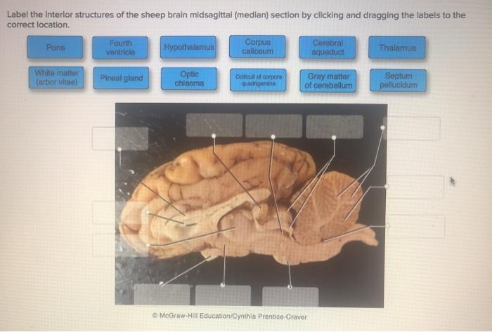
Solved Label the interior structures of the sheep brain ...
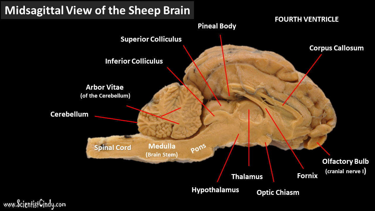
PDF Sheep Brain Midsagittal Section - Dr. Scott Croes' Website Sheep Brain Frontal Section 19 20 21 16 18 18 19 Note: Together the globus pallidus and putamen are referred to as the lentiform nucleus

Sheep Brain Dissection – Krysta H, Rinda G, Keira N, Cindy M ...
Sheep Brain Neuroanatomy Online Self-Test | KPU.ca ... Sheep Brain Neuroanatomy Online Self-Test. Use each diagram as a reference, and selected the correct answer for each lettered structure. You may find it useful to open the diagrams in a separate window to review while answering each question. Dorsal Surface.
Physiological Psychology
Functions - Sheep Brain Dissection Hypothalamus- is a portion of the brain that contains a number of small nuclei with a variety of functions. One of the most important functions of the hypothalamus is to link the nervous system to the endocrine system via the pituitary gland. Cerebral Acquesduct-a fluid-filled canal that runs through the midbrain connecting the third and fourth ...
11c Brain Anatomy
PDF Sheep Neuroanatomy Lab- Labeling Worksheet Psychology 2315 ... Sheep Neuroanatomy Lab- Labeling Worksheet Psychology 2315- Brain and Behaviour Kwantlen Polytechnic University Figure 1: Dorsal view Cerebellum, Frontal lobe, Occipital lobe, Parietal lobe, and Temporal lobe. Temporal Parietal Lobe Frontal Lobe Cerebellum Occipital Lobe

Lateral View of Brain, Labeled
BIO201-Sheep Brain

Sheep Brain Images

SCB209 - Lab2 - Natural Sciences Open Educational Resources
Sheep Brain Dissection with Labeled Images
Sheep Brain Explora on Guide
Sheep Brain Dissection Bi - BIOLOGY JUNCTION
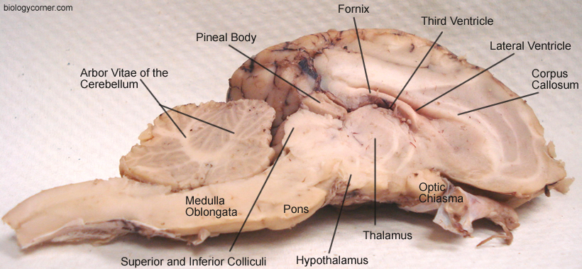
Sheep Brain Dissection with Labeled Images

BIOL 160: Human Anatomy and Physiology
Mapping The Creative Brain - Sheep Brain Labeled Upper View ...

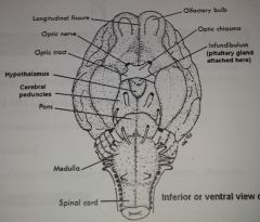
Diagram of Sheep Brain - Inferior view

Resources for Teaching Mammalian Neuroanatomy Using Sheep ...
Lab Exam 3: Anatomy of Sheep Brain; Histology Flashcards ...
Sheep brain dissection - Bisc 163 - StuDocu

Labeling- Sheep brain Figure 17.11 intact sheep brain Diagram ...
Labeled Superior View Of Real Brain, HD Png Download - kindpng
Solved art-labeing activity: midsagittao section of the ...

Sheep brain | Atlas of Comparative Vertebrate Anatomy

Comparative Anatomy - an overview | ScienceDirect Topics
0 Response to "37 sheep brain diagram labeled"
Post a Comment