39 diagram of facial muscles
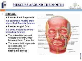
called the kissing muscle because it causes the lips to close and pucker. Platysma. pulls the lower lip and corner of the mouth sideways and down to change the facial expression. It also depresses and wrinkles the skin of the neck, a common sign of aging, and helps in the lowering of the mandible. Cranial Aponeurosis. Identifying Facial Muscles that Need Shortening. Muscles that need shortening include: The Platysma along the sides of the neck and jowls which help tighten the neck and jaw area. The Depressor Anguli Oris which helps lift the nasal folds to the side of the mouth and nose. The Levator Superioris Nasi, Levator Superioris and Zygomaticus Major ...
The muscles of the face overlap and crisscross over each other, creating a mask of muscle over the skull and jawbone. They attach to various parts of the skull and other muscles, allowing for a ...

Diagram of facial muscles
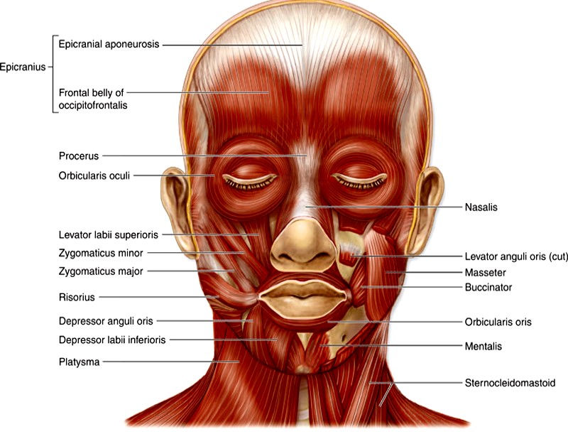
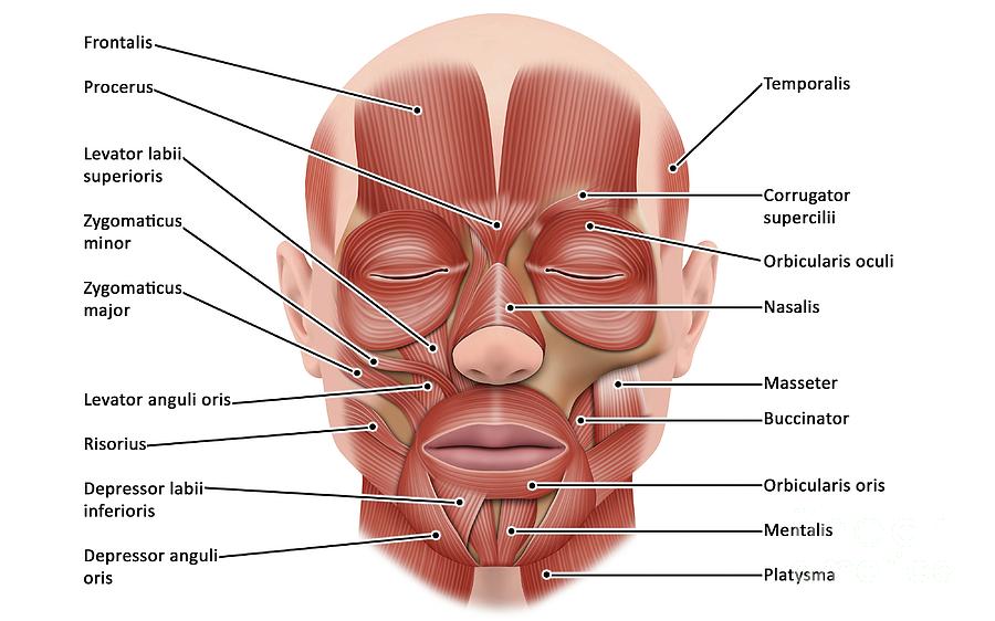
The muscles of facial expression are located in the subcutaneous tissue, originating from bone or fascia, and inserting onto the skin. By contracting, the muscles pull on the skin and exert their effects. They are the only group of muscles that insert into skin. Oct 28, 2021 · Face muscle anatomy. Found situated around openings like the mouth, eyes and nose or stretched across the skull and neck, the facial muscles are a group of around 20 skeletal muscles which lie underneath the facial skin. The majority originate from the skull or fibrous structures, and connect to the skin through an elastic tendon. The face has many muscles, each with its own unique function. Some, but not all, are controlled by CN-VII. These muscles are known as "the muscles of facial expression". Unlike other muscles, the facial muscles insert directly into the skin. Contraction of the muscles causes the skin to move.
Diagram of facial muscles. Feb 7, 2020 - Explore Susan Torrey's board "face diagrams" on Pinterest. See more ideas about facial anatomy, facial aesthetics, face anatomy. Facial muscles include a group of 20 flat skeletal muscles, which reside under flat facial skin. They originate from the skull and fibrous structures and use an elastic tendon in order to radiate to the skin. These muscles are unique in the way that they are positioned surrounding facial openings, such as the ears, eyes, nose, and mouth, while ... Your facial muscles work together to control the parts of your face. They are essential to chewing, facial expressions and other functions. Weakness or paralysis of your face muscles can be a temporary condition or a serious medical problem. See a healthcare provider right away if you have facial palsy or any trouble smiling, talking or eating. Muscles of the Head and Neck. Humans have well-developed muscles in the face that permit a large variety of facial expressions. Because the muscles are used to show surprise, disgust, anger, fear, and other emotions, they are an important means of nonverbal communication. Muscles of facial expression include frontalis, orbicularis oris, laris ...
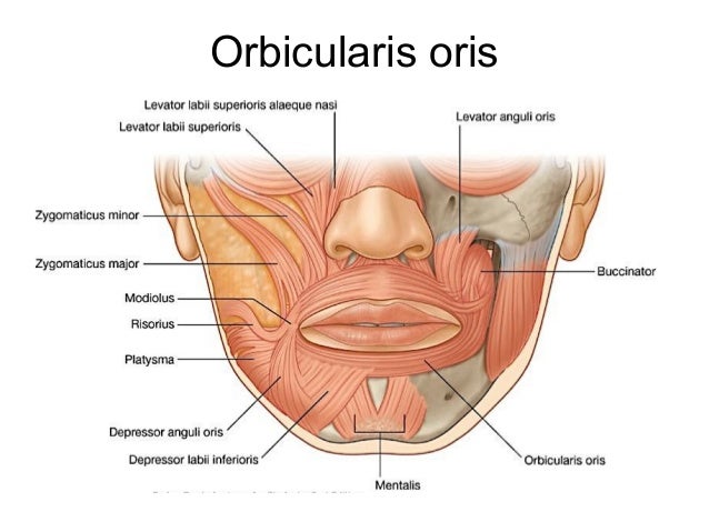
Function: This facial muscle helps to hold food inside the mouth in proper position and aids in chewing. Flattening the cheeks and pulling the angle of the mouth backwards is supported by this muscle. #5. Mentalis Muscle of the Face: The furrow between the lower lip and chin is formed by this muscle of the face. In other words, it can be said that this facial muscle is located at the tip of ... Temporalis, Sternocleidomastoid, Orbicularis Oris, Orbicularis Oculi, Masseter, Zygomaticus, Mentalis, Frontalis, Corrugator, Buccinator, Trapezius, Risorius. Facial muscles (Musculi faciales) The facial muscles, also called craniofacial muscles, are a group of about 20 flat skeletal muscles lying underneath the skin of the face and Most of them originate from the bones or fibrous structures of the skull and radiate to insert on the. Contrary to the other skeletal muscles they are not surrounded by a fascia, with the exception of the ... Important Skeletal Muscles, 286 Muscles of Facial Expression, 287 Muscles of Mastication, 288 Muscles That Move the Head, 288 Trunk Muscles, 289 Muscles of the Thorax, 289 Muscles of the Abdominal Wall, 289 Muscles of the Back, 290 Muscles of the Pelvic Floor, 290 Upper Limb Muscles, 293 Muscles Acting on the Shoulder Girdle, 293
Dec 30, 2010 · Posted December 30, 2010 in BOTOX® Cosmetic and Dysport®, Face. In order to understand what to expect from Botox or Dyport treatment, a basic knowledge of muscle anatomy really helps. Look at the diagram below: Anatomy diagram: A. Frontalis muscle: contraction raises the eyebrows and causes horizontal brow wrinkles. Injection of Botox or ... Facial anatomy at-a-glance. Dec 06, 2017. Knowing the facial anatomy is fundamental to performing more than aesthetic surgery. A provider's lack of understanding of the intricate web of facial muscles, nerves, arteries and more can turn a relatively simple injection technique, with botulinum toxin or a filler, into a serious complication. The muscles of the head and neck perform many important tasks, including movement of the head and neck, chewing and swallowing, speech, facial expressions, and movement of the eyes. These diverse tasks require both strong, forceful movements and some of the fastest, finest, and most delicate adjustments in the entire human body. Doing facial exercises, or facial yoga, is a natural way to make your face look younger by firming muscles and reducing wrinkles. These are also good exercises to do if you have a muscle problem on your face, creating stronger muscles for a toned and more confident look.
Diagram of facial muscles, front view (Photos Framed Prints ...
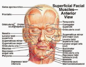
Jun 4, 2015 - Muscles of the face - superficial facial muscles - human anatomy diagram : perform many important tasks, including movement of the head and neck, chewing..

Muscles of the Face - Mimetic Muscles - Medical Art Library
Muscle Charts of the Human Body For your reference value these charts show the major superficial and deep muscles of the human body. Superficial and deep anterior muscles of upper body

Facial Muscles Stock Illustrations – 473 Facial Muscles Stock ...
1 4 9 3 2 10 4 7 11 6 8 5 12 13 Muscles of Facial Expression Blood Supply: External Carotid Artery Motor Innervation: Facial Nerve (Vll) Sensory Innervation: Trigeminal Nerve (V) 1) Frontalis (worry muscle): a. Actions: Raises eyebrows, furrows brow b. Innervation: Facial Nerve (Vll) c. Origin: from galea aponeurotica d. Insertion: to skin above the eyebrows 2) Occipitalis – not shown on ...
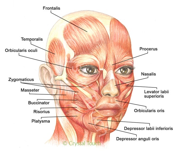
Our facial muscles and their functions • Crystal Touch Bell's ...
Start studying Facial Muscles. Learn vocabulary, terms, and more with flashcards, games, and other study tools.
Muscles of facial expression
Start studying Facial Muscles. Learn vocabulary, terms, and more with flashcards, games, and other study tools.

Facial Muscle Chart
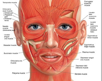
Anatomy note Odysee Channel, Please Subscribe to Support. Human facial muscle diagram In this image, you will find galea aponeurotica, frontalis, corrugator, levator labii superiors alaeque nasi, levator superons, obicularis oris, risonus, mentalis, platysma in it.

Facial Muscles and Expressions - Classic Human Anatomy in ...
The named muscle is highlighted in green and the arrows show the direction of muscle movement. Diagram of the Facial Muscles Key to facial muscles Procerus Corrugator Depressor Supercilii Orbicularis Oculi (inner ring = pretarsal portion) Zygomaticus Major Levator Labii Superioris Masseter Levator Anguli Oris Buccinator Risorius

Facial Muscles Of The Female Stock Illustration - Download ...
Simply put, this Facial Muscle Chart shows the muscle structure of the face and help boost confidence in your MyoLift session. For beginners, people often wonder where to apply the microcurrent applicators.It is makes a big difference in your microcurrent facial results when you understand how the muscles are positioned on the face and neck.

Human Face Anatomy Diagram | Muscles of the face, Human body ...
Here are a number of highest rated Human Anatomy Face Muscles Diagram pictures on internet. We identified it from honorable source. Its submitted by dispensation in the best field. We put up with this kind of Human Anatomy Face Muscles Diagram graphic could possibly be the most trending subject next we portion it in google gain or facebook.

Human facial muscle diagram
In this article, we will discuss the motor points of important facial muscles with a clear diagram. So, let's get started. Indications for facial muscle stimulation. The only indication of face muscle stimulation is paralysis/paresis of facial muscle. There are few known conditions where paresis of face muscle occurs due to partial or ...

Head And Neck Muscles Labeled Anatomical Diagram, Facial ...
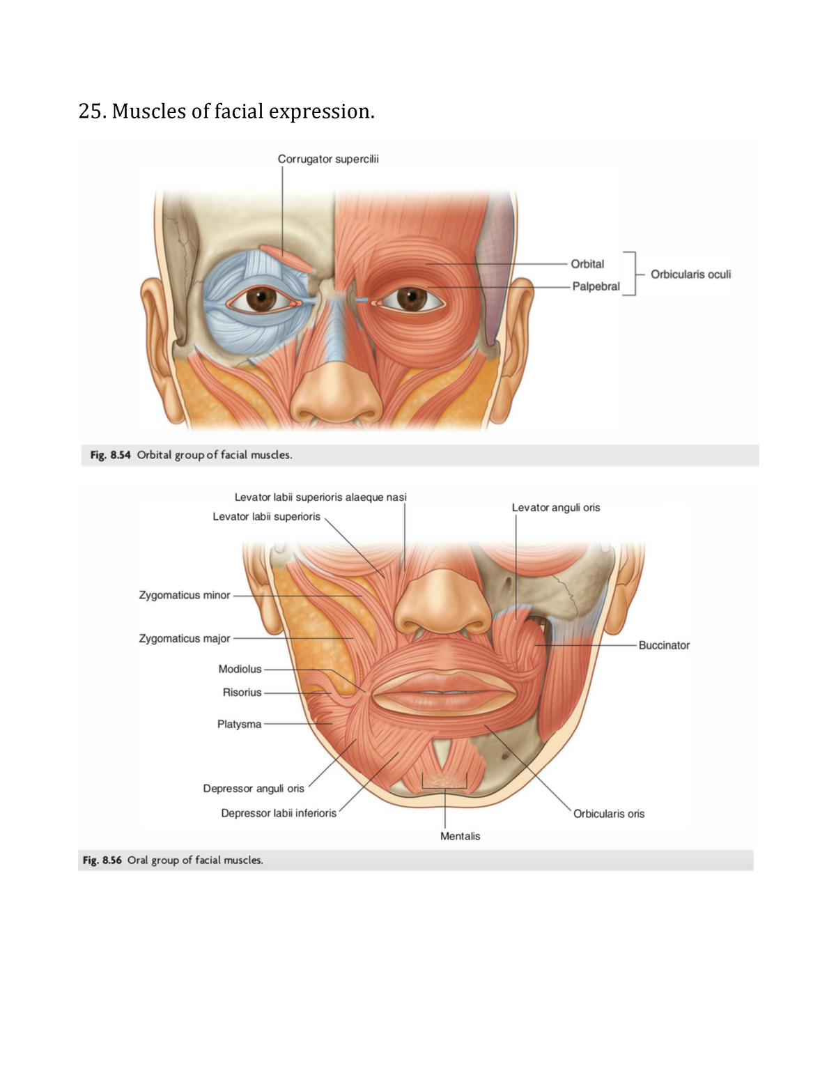
The muscles of facial expression (also known as the mimetic muscles) can generally be divided into three main functional categories: orbital, nasal and oral. These muscles are all innervated by the facial nerve (CN VII).¹. These striated muscles broadly originate from the surface of the skull and insert onto facial skin.

Muscles of Chewing and Swallowing - Physiology - AmeriCorps ...
The facial nerve is also known as the seventh cranial nerve (CN7). This nerve performs two major functions. It conveys some sensory information from the tongue and the interior of the mouth.

Facial Muscles Front View Diagram | Quizlet
The face has many muscles, each with its own unique function. Some, but not all, are controlled by CN-VII. These muscles are known as "the muscles of facial expression". Unlike other muscles, the facial muscles insert directly into the skin. Contraction of the muscles causes the skin to move.

Anatomy of the Head and Neck - Medical Illustrations showing ...
Oct 28, 2021 · Face muscle anatomy. Found situated around openings like the mouth, eyes and nose or stretched across the skull and neck, the facial muscles are a group of around 20 skeletal muscles which lie underneath the facial skin. The majority originate from the skull or fibrous structures, and connect to the skin through an elastic tendon.

Facial Muscles Face Anatomy Facial Nerve, PNG, 1024x879px ...
The muscles of facial expression are located in the subcutaneous tissue, originating from bone or fascia, and inserting onto the skin. By contracting, the muscles pull on the skin and exert their effects. They are the only group of muscles that insert into skin.

Zbrushtuts - Anatomy of Facial Expression Facial anatomy for ...

Axial Muscles of the Head, Neck, and Back | Anatomy and ...
Facial Muscles | Etsy

facial muscles side view Diagram | Quizlet

Facial Muscles. Graphic Illustration. Hand Drawing, Contour ...

Muscles of facial expression

FACIAL MUSCLE Human Face Anatomy Detailed Axial Muscle POSTER ...
Face - Muscles - AnatomyQA

Anatomy of the facial muscles. Reprinted under Creative ...
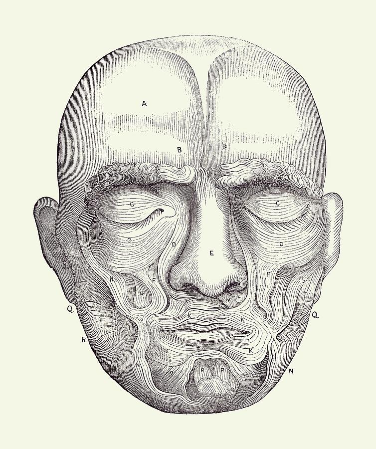
Facial Muscles - Simple Diagram - Vintage Anatomy Print 2 by Vintage Anatomy Prints
Face Muscular Diagram 2 - Vintage Anatomy 2 by Vintage Anatomy Prints

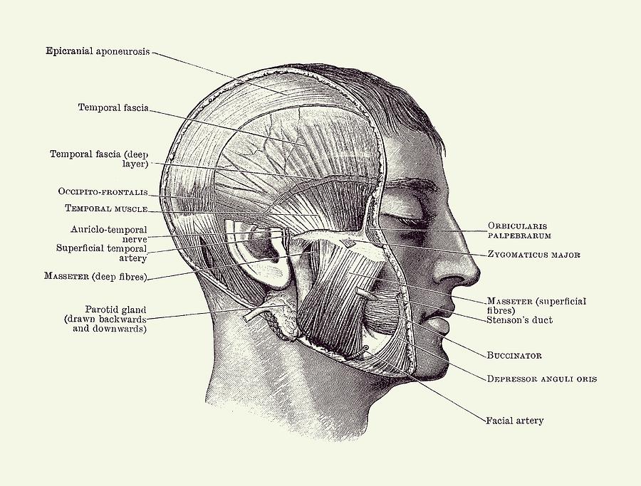
Ch 10- Lateral view of Muscles of the Scalp, Face, and Neck ...
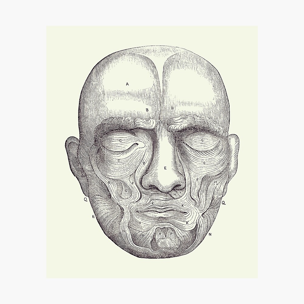
Facial Muscles - Simple Diagram - Vintage Anatomy Print 2 ...

Facial Muscles | Etsy
Facial muscles function, anatomy, arteries, veins, names ...
All muscles - Summary Gray's Anatomy for Students - 25 ...

Pin by Emily Bowen on Health | Muscles of the face, Facial ...

Anatomy of Facial Expression - Paperback (English)
Essential anatomy for facial injections

Free Anatomy Quiz - The Muscles of the Face, Locations - Quiz 1

Facial muscles Diagram | Quizlet

5: Upper face muscles used to produce facial expressions ...
Face Muscle Anatomy Photograph by Maurizio De Angelis/science ...
Face muscles - Teaching resources
0 Response to "39 diagram of facial muscles"
Post a Comment