37 pseudostratified columnar epithelium diagram
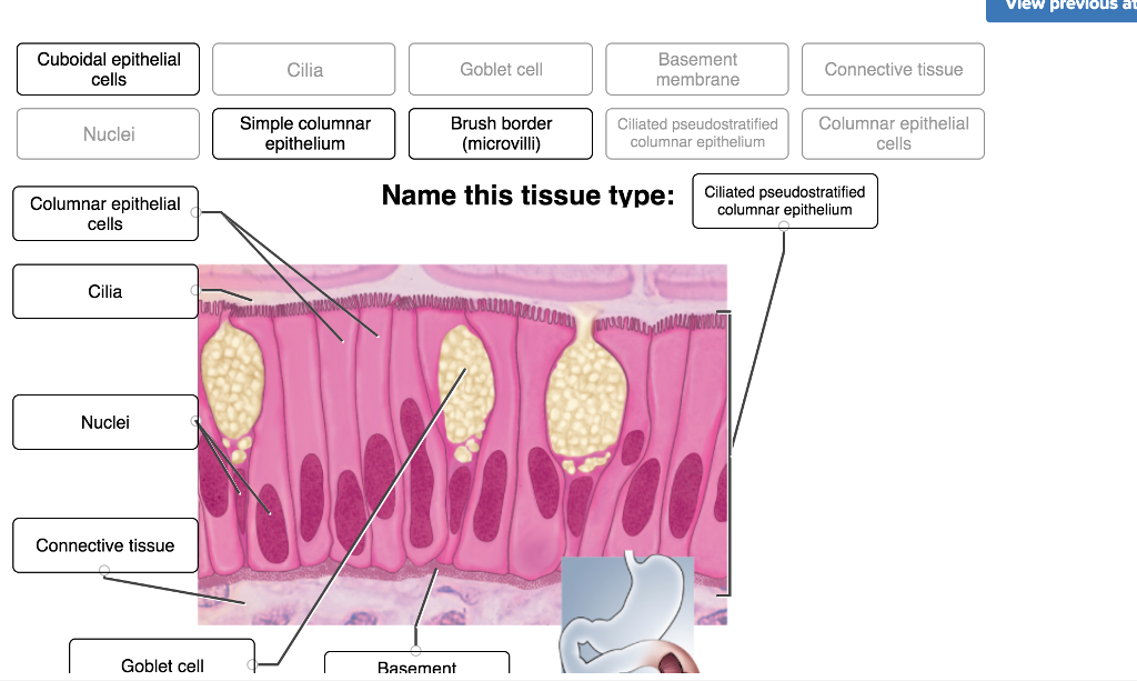
Simple columnar epithelium- structure, functions, examples The simple columnar epithelium is a type of epithelium that is formed of a single layer of long, elongated cells mostly in areas where absorption and secretion are the main functions. Like cuboidal epithelium, the cells in the columnar epithelium are also modified to suit the function and structure of the organ better. Pseudostratified ciliated epithelium Pseudostratified ciliated epithelium (40X) Human respiratory tract The bar in this image shows the thickness of the layer of ciliated pseudostratified epithelium. The rest of the tissue below the epithelium is mostly connective tissue, but there are some mucous glands (glandular epithelium) just below the surface.
Holocrine - Wikipedia Holocrine is a term used to classify the mode of secretion in exocrine glands in the study of histology.Holocrine secretions are produced in the cytoplasm of the cell and released by the rupture of the plasma membrane, which destroys the cell and results in the secretion of the product into the lumen.. Holocrine gland secretion is the most damaging (to the cell itself and …

Pseudostratified columnar epithelium diagram
Pseudostratified Columnar Epithelium Function & Location ... The pseudostratified columnar epithelium is present on the internal surface of the upper respiratory tract, including nasal passages and the lower respiratory tract, including the trachea and the... Pseudostratified Columnar Epithelium - Kit Ng, Ph.D. Pseudostratified columnar epithelium gets its name because while the cells are columnar in shape, their nuclei do not arrange at the bottom (or basal side) of the cell. Rather the nuclei are arranged in an almost random fashion. However, they are true simple epithelium because it is only a single layer of cells, and the… en.wikipedia.org › wiki › ApocrineApocrine - Wikipedia Apocrine (/ ˈ æ p ə k r ɪ n /) is a term used to classify exocrine glands in the study of histology.Cells which are classified as apocrine bud their secretions off through the plasma membrane producing extracellular membrane-bound vesicles.
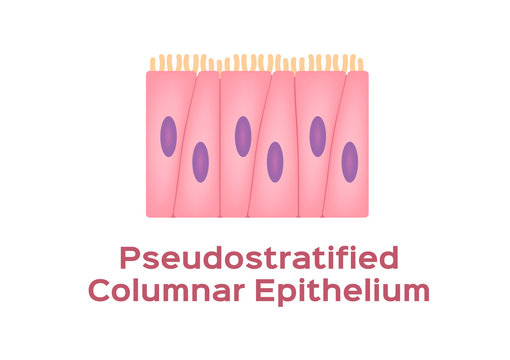
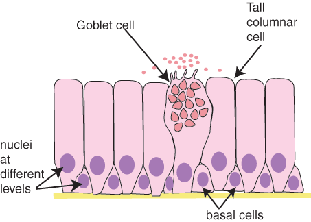
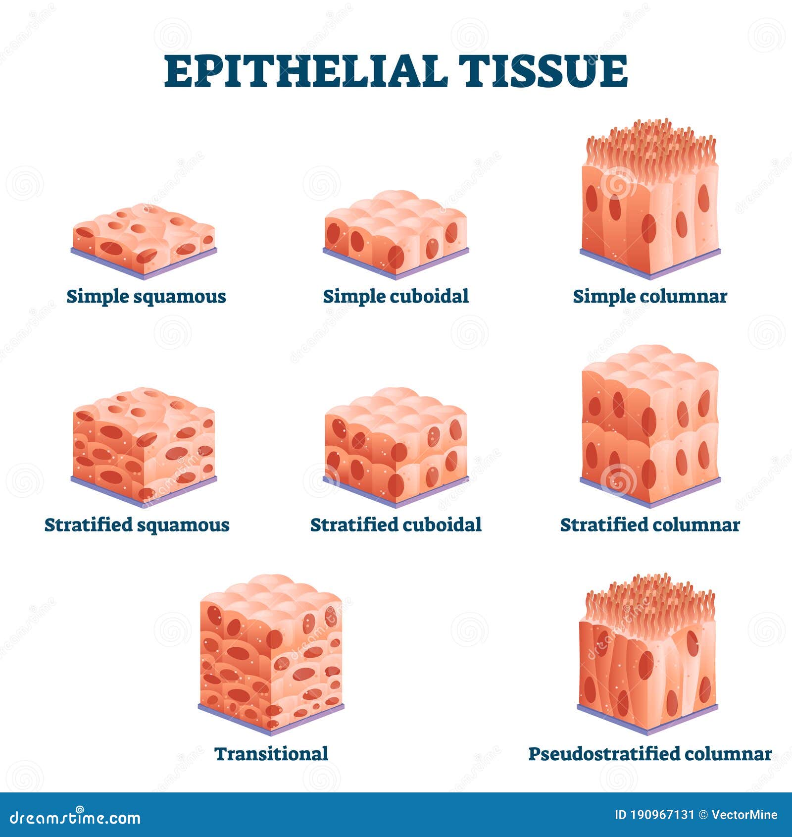
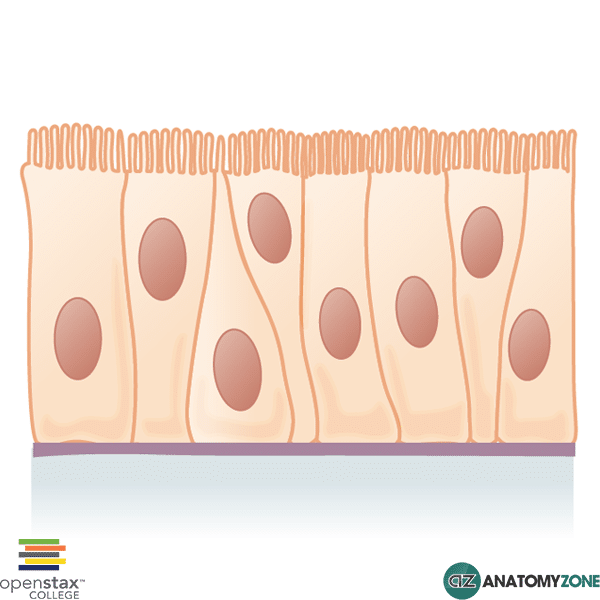
Pseudostratified columnar epithelium diagram. Pseudostratified epithelium - Eugraph In a pseudostratified epithelium one can see nuclei n at several different levels, giving an appearance of an epithelium composed of layers of cells (a stratified epithelium).. However it has been shown that all of the cells of a pseudostratified epithelium are in contact with the basement membrane bm. The term pseudo (false) reminds one that this is not a stratified epithelium. Pseudostratified Columnar Epithelium | Histology, Anatomy ... Diagrammatic Illustration Of The Pseudostratified Columnar Epithelium (Source: dartmouth.edu) The diagram above illustrates a pseudostratified epithelium. In terms of shape, the cells are variously shaped but most of them are taller than wide (as implied by their name, columnar) and are shown to appear like miniature pillars. Ciliated Epithelium - Concept, Structure, Function and ... Pseudostratified Ciliated Epithelium. Pseudostratified ciliated columnar epithelia are tissues that are made up of only one layer of cells but appear to be made up of numerous layers when viewed in cross-section. The nuclei of epithelial cells are at extremely different levels, giving the appearance of stratification. Pseudostratified Columnar Epithelium - Definition & Function Pseudostratified Columnar Epithelium Definition. Pseudostratified columnar epithelia are tissues formed by a single layer of cells that give the appearance of being made from multiple layers, especially when seen in cross section. The nuclei of these epithelial cells are at different levels leading to the illusion of being stratified.
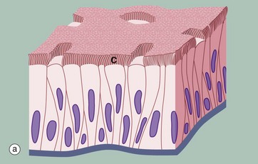
Stratified cuboidal epithelium- structure, functions, examples Cell Organelles- Definition, Structure, Functions, Diagram; Pseudostratified columnar epithelium- structure, functions, examples; Transitional epithelium- definition, structure, functions, examples; Simple squamous epithelium- structure, functions, examples; Epithelial vs Connective tissue- Definition, 15 Differences, Examples ; Subscribe us to receive latest notes. … Pseudostratified columnar epithelium- structure, functions ... The pseudostratified columnar epithelium is a type of epithelium consisting of a single layer of cells that gives the appearance of being multiple layers because the nuclei of the cells are present at different levels. This epithelium is histologically a simple epithelium even though in a crosssection, it might appear as a stratified epithelium. Pseudostratified Columnar Epithelium - AnatomyZone This diagram represents pseudostratified columnar epithelium. This is a special type of simple epithelium called pseudostratified epithelium as it resembles stratified epithelium due to the positioning of the cellular nuclei, but is comprised of only a single layer of cells. Chapter 2, Page 2 - HistologyOLM - SteveGallik.org Introduction Diagram of a pseudostratified columnar epithelium A pseudostratified columnar epithelium is a special type of simple epithelium that consists of a falsely-stratified single layer of epithelial cells resting on a basement membrane. Many, if not most, of the cells, are columnar.
Simple Cuboidal Epithelium Function & Location | What Is ... 13.12.2021 · Simple Cuboidal Epithelium: Labeled Diagram Simple cuboidal epithelial cells are shaped like cubes, and the nucleus of each cell is large and located close to the center of the cell. Pseudostratified Columnar Epithelium: Location & Function ... Pseudostratified columnar epithelium is tissue composed of a single layer of columnar cells that line the space of an organ cavity or vessel. It is particularly useful in the secretion and ... Epididymis Histology Slide and Identification Points with ... The pseudostratified columnar epithelium (with a variety of stereocilia) is also shown on the epididymis labeled diagram. A circularly arranged smooth muscle fibers that surround the epididymal tubules are also identified in the labeled diagram. There is a clump of spermatozoa present in the lumen of the epididymis. Pseudostratified Ciliated Columnar Epithelium Unlike the epithelium of the skin, a pseudostratified ciliated columnar epithelium appears to have multiple layers, but is actually only comprised of a single sheet of cells. The positioning of the nuclei within the individual columnar cells causes this illusion. These structures, which are easily identifiable with the help of a microscope, are ...
Nasopharynx Definition, Anatomy, Function, Diagram 16.02.2018 · The nasopharynx is primarily lined by two types of epithelia, with the stratified squamous epithelium comprising around 60% of its inner walls [5].The nasopharynx is also the only section of the pharynx to have pseudostratified columnar respiratory epithelium [6], the specialized epithelium (ciliated and containing goblet cells) for the respiratory tract [7].
Solved Label the following Tissue in your drawing Diagram ... Transcribed image text: Label the following Tissue in your drawing Diagram Magnification: Basement membrane Principal cells Epididymis Pseudostratified columnar epithelium Basal cell Spermatozoa Tissue Label the following in your drawing Diagram Magnification: Seminal vesicles Pseudostratified columnar epithelium Lumen Basal cells 1. Compare and contrast the functions of the epididymis and ...
PseudoStratified columnar epithelium | Psychology notes ... Pseudostratified epithelium is also sometimes referred to as respiratory epithelium, since ciliated pseudostratified columnar epithelia is mainly found in the larger respiratory airways of the nasal cavity, trachea and bronchi. Find this Pin and more on File Diagrams by Histopedia. Psychology Notes. Nasal Cavity.
PseudoStratified columnar epithelium | Psychology notes ... Pseudostratified epithelium is also sometimes referred to as respiratory epithelium, since ciliated pseudostratified columnar epithelia is mainly found in the larger respiratory airways of the nasal cavity, trachea and bronchi. Find this Pin and more on File Diagrams by Histopedia. Histology Slides. Psychology Notes.
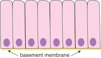
Simple squamous epithelium- structure, functions, examples 27.09.2020 · Simple squamous epithelium is a type of simple epithelium that is formed by a single layer of cells on a basement membrane. It is a type of epithelium formed by a single layer of squamous or flat cells present on a thin extracellular layer, called the basement membrane. This epithelium is also termed the pavement epithelium because the cells appear like tiles on …
Pseudostratified Columnar Epithelium Diagram | Quizlet Label the parts of the pseudostratified columnar epithelium. Learn with flashcards, games, and more — for free.
Simple Columnar Epithelium Labeled Diagram This type of epithelium is adapted for secretion and/or absorption, and can also be protective. Simple secretory columnar epithelium lines the stomach and uterine schematron.org simple columnar epithelium that lines the intestine also contains a few goblet cells. Simple Columnar Epithelium: A Labeled Diagram and Functions Epithelium is a tissue ...
Chapter 2, Page 4 - HistologyOLM Introduction Diagram of a pseudostratified columnar epithelium A nonciliated pseudostratified columnar epithelium is a specialized form of pseudostratified columnar epithelium that lines the epididymis of the male reproductive system.
Simple Columnar Epithelium: A Labeled Diagram and ... Epithelium is a tissue that lines the internal surface of the body, as well as the internal organs. Simple epithelium is one of the types of epithelium that is divided into simple columnar epithelium, simple squamous epithelium, and simple cuboidal epithelium. Bodytomy provides a labeled diagram to help you understand the structure and function of simple columnar epithelium.
Pseudostratified columnar epithelium Diagram | Quizlet Start studying Pseudostratified columnar epithelium. Learn vocabulary, terms, and more with flashcards, games, and other study tools.
pseudostratified ciliated columnar epithelium | Tissue ... Feb 24, 2016 - pseudostratified ciliated columnar epithelium diagram - Google Search
Merocrine - Wikipedia Merocrine (or eccrine) is a term used to classify exocrine glands and their secretions in the study of histology.A cell is classified as merocrine if the secretions of that cell are excreted via exocytosis from secretory cells into an epithelial-walled duct or ducts and then onto a bodily surface or into the lumen.. Merocrine is the most common manner of secretion.
Pseudostratified Columnar Epithelium Histology - Jotscroll The Pseudostratified Columnar Epithelium lining the ductus epididymis and certain other parts of the male reproductive tract does not have cilia or goblet cells. Well labeled diagram of Pseudostratified columnar epithelium showing the cilia, goblet cells, lamina propria, basement membrane and epithelial cells
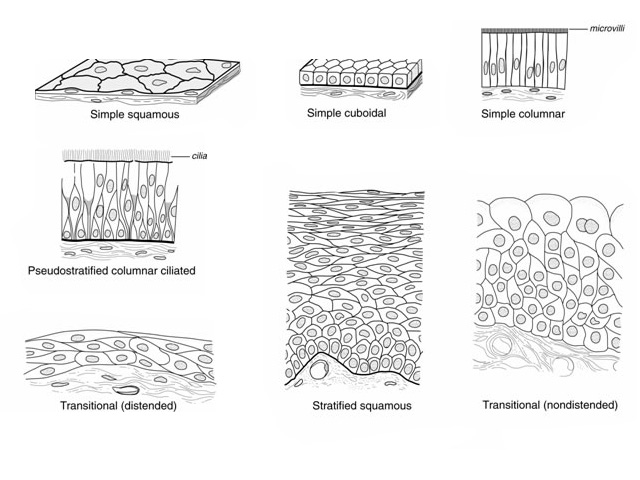
Epithelial Tissue: Structure with Diagram, Function, Types ... Pseudostratified Columnar: Respiratory passage and ducts of many glands: Similar to columnar epithelium but all the cells are not of similar height: Protection, secretion and movement of mucous: Transitional epithelia or urothelium: Urinary bladder, urethra, ureter: Stratified epithelium, which can contract or expand as per the requirement. Cells are cuboidal …
[Solved] PSEUDOSTRATIFIED CILIATED COLUMNAR EPITHELIUM ... In the above diagram all parts are labelled such as , Pseudostratified columnar epithelium cells, ciliated border, and goblet cells . 2 - Location of the pseudostratified columnar epithelium is trachea and upper respiratory tract, in male it is located in the epididymis, prostate gland and vas deference, and in females it is located in the ...
Epithelial Tissue | histology A. Simple columnar epithelium. Slide 29 (small intestine) View Virtual Slide. Slide 176 40x (colon, H&E) View Virtual Slide. Remember that epithelia line or cover surfaces. In slide 29 and slide 176, this type of epithelium lines the luminal (mucosal) surface of the small and large intestines, respectively. Refer to the diagram at the end of this chapter for the tissue …
Simple epithelium: Location, function, structure | Kenhub Pseudostratified columnar epithelium with stereocilia, another sub-classification, can be found in the epididymis, a highly coiled genital duct of the male. Non-ciliated pseudostratified columnar epithelia can be found in the membranous part of male vas deferens.
Lab Quiz # 3 Diagram | Quizlet pseudostratified columnar epithelium. single layer of flattened cells. simple squamous epithelium. Single row of elongated cells, but some cells don't reach the free surface. pseudostratified columnar epithelium. forms walls of capillaries and air sacs of lungs. simple squamous epithelium. Provides lining of urethra of males and parts of pharynx . stratified …
4.2 Epithelial Tissue - Anatomy & Physiology Pseudostratified columnar epithelium is a type of epithelium that appears to be stratified but instead consists of a single layer of irregularly shaped and differently sized columnar cells. In pseudostratified epithelium, nuclei of neighboring cells appear at different levels rather than clustered in the basal end. The arrangement gives the ...
en.wikipedia.org › wiki › ApocrineApocrine - Wikipedia Apocrine (/ ˈ æ p ə k r ɪ n /) is a term used to classify exocrine glands in the study of histology.Cells which are classified as apocrine bud their secretions off through the plasma membrane producing extracellular membrane-bound vesicles.
Pseudostratified Columnar Epithelium - Kit Ng, Ph.D. Pseudostratified columnar epithelium gets its name because while the cells are columnar in shape, their nuclei do not arrange at the bottom (or basal side) of the cell. Rather the nuclei are arranged in an almost random fashion. However, they are true simple epithelium because it is only a single layer of cells, and the…
Pseudostratified Columnar Epithelium Function & Location ... The pseudostratified columnar epithelium is present on the internal surface of the upper respiratory tract, including nasal passages and the lower respiratory tract, including the trachea and the...










0 Response to "37 pseudostratified columnar epithelium diagram"
Post a Comment