38 upper airway anatomy diagram
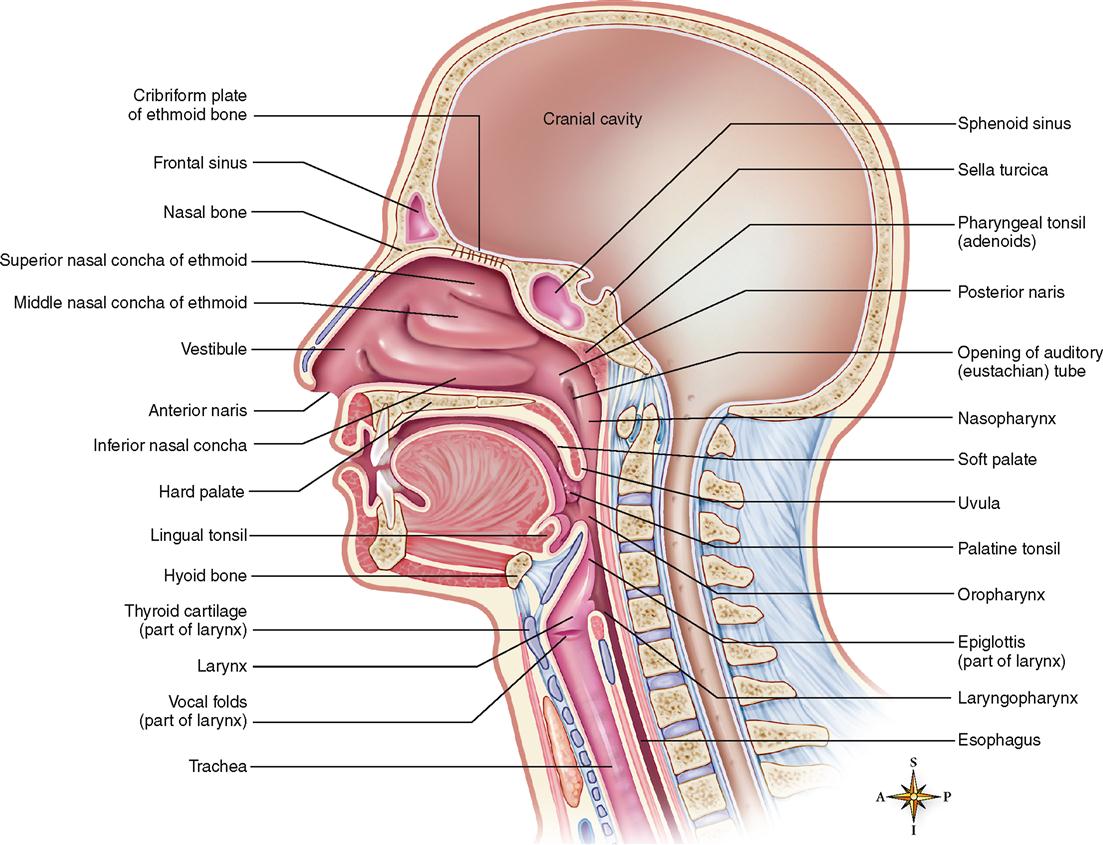
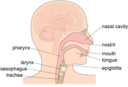
Upper Airway Obstruction - StatPearls - NCBI Bookshelf The upper airway consists of the nasal cavities, oral cavity, pharynx, and larynx. The pharynx is further subdivided into the nasopharynx, oropharynx, and hypopharynx. The larynx is divided into three regions, dependent on their relationship to the vocal cords (glottis). These areas are the supraglottis, glottis, and subglottis. The supraglottis consists of the epiglottis, the arytenoids, the ... 👉 Throat Anatomy (February 2022) Jan 12, 2022 · Epiglottis-This is the part that closes the airway during swallowing. It is simply a piece of cartilage that is behind the tongue and at the start of the larynx. (1, 2, 5) Picture 9 : The trachea is in comparison to the larynx and the lungs. Image Source : upload.wikimedia.org. In this one of its most important functions is to protect the airway.
Anatomy of the upper airway - ICST This diagram outlines the main anatomical structures of the upper airway that are important when inserting a tracheostomy tube. Anatomy of the upper airway The tracheostomy is the procedure of creating an artificial opening into the front of the trachea, to bypass the airway superior to this where there may be a cancer, obstruction, or to ...

Upper airway anatomy diagram
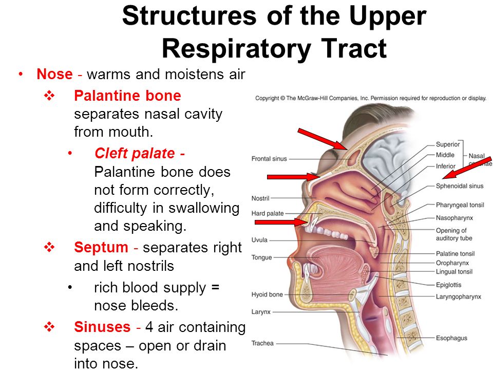
EMT Anatomy - Airway Diagram | Quizlet Start studying EMT Anatomy - Airway. Learn vocabulary, terms, and more with flashcards, games, and other study tools. PDF 1 Airway Anatomy - Notes For ANZCA Primary Exam 1.Airway Anatomy - 2 Basic Structural Anatomy of Upper Airway & Larynx Laryngeal Skeleton • Hyoid: o Attachement for thyroid cartilage via throhyoid membrane & extrinsinc mm of larynx o U shaped body o Body & paired greater & lesser horns • Skeleton has 9 cartilges: o Singles: ! Thyroid ! Cricoid Anatomy and Physiology: The Upper Respiratory System The nasal cavity is a chamber of the internal nose that connects to the nasopharynx. Air is inhaled through the nostrils and warmed as it passes through the nasal cavities. There is a mucous membrane that lines the cavity; the mucus helps trap unwanted particles in inhaled air. Ethmoid and Other Skeletal Structures
Upper airway anatomy diagram. Structure and function of the upper airways | Deranged ... The upper airway is full of muscles, the anatomy of which is the nightmarish province of ENT and maxillofacial surgeons. For the intensivist, it will likely suffice to know that their function in respiration is basically to not collapse. And to collapse, apparently, is their natural tendency. Respiratory System Anatomy, Diagram & Function | Healthline Upper respiratory tract: Composed of the nose, the pharynx, and the larynx, the organs of the upper respiratory tract are located outside the chest cavity. Nasal cavity: Inside the nose, the ... Anatomy, Airway - StatPearls - NCBI Bookshelf The muscles of the pharynx and larynx provide the structure of the upper airways and form from striated muscles under visceral and somatic control. They relate to the action of swallowing. The lower airways have a layer of smooth muscle within their walls. Nose Anatomy, Function & Diagram | Body Maps Jan 21, 2018 · The nose is the body's primary organ of smell and also functions as part of the body's respiratory system. Air comes into the body through the nose. As it passes over the specialized cells of the ...
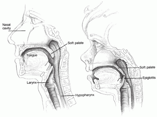
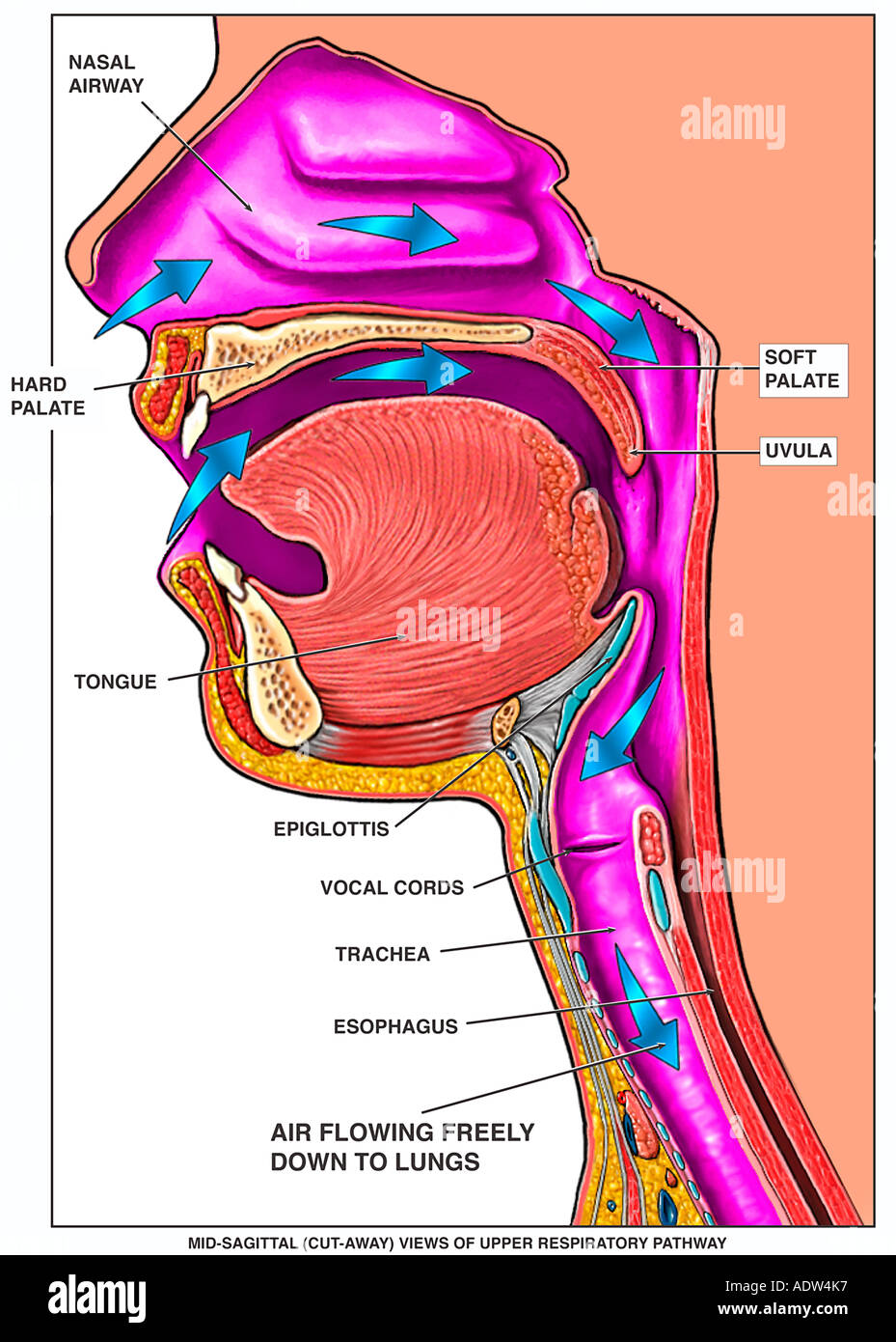
Anatomy and Physiology (A&P) of the Upper Airway | First ... The epiglottis is one of the main anatomic landmarks in the airway. It looks like a floppy piece of cartilage covered by mucosa-which is exactly what it is-and it feels like the tragus- the cartilage at the opening of the car canal. The epiglottis is attached to the hyoid and thence to the mandible by a series of ligaments and muscles. Upper Respiratory System - SmartDraw Upper Respiratory System. Create healthcare diagrams like this example called Upper Respiratory System in minutes with SmartDraw. SmartDraw includes 1000s of professional healthcare and anatomy chart templates that you can modify and make your own. 21/22 EXAMPLES. EDIT THIS EXAMPLE. Stages of swallowing: Deglutition - Kenhub Mar 20, 2022 · The oral phase of swallowing is the first stage of deglutition, and it is a voluntary process. It is also commonly known as the buccal phase.It involves the contraction of the tongue to push the bolus up against the soft palate and then posteriorly into the oropharynx by both the tongue and the soft palate. PDF Advanced Airway Management for EMTs (2).ppt Objectives Review anatomy relevant to airway managementReview anatomy relevant to airway management Relate key differences in airway structures and how they influence successful bag mask ventilation (BMV) Describe the process of opening the airway and maitii itintaining it Describe the indications, limitations, proper sizing and contraindications of BLS airwaysizing, and contraindications of ...
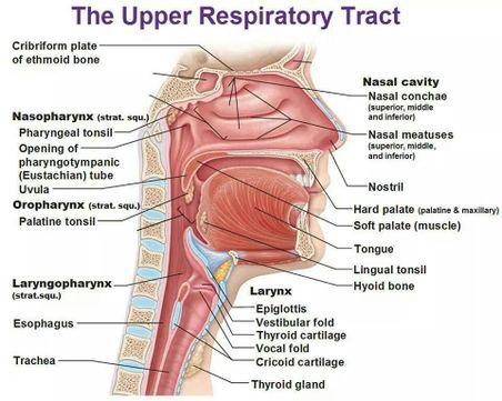
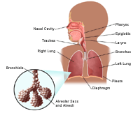
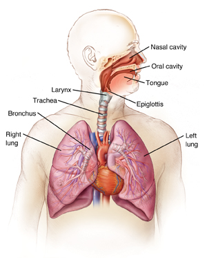
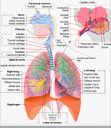
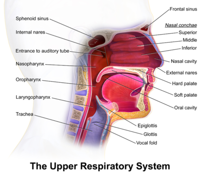
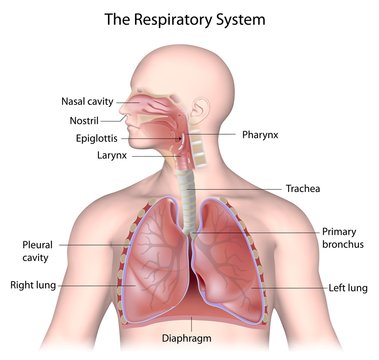
Respiratory system - Wikipedia In humans and other mammals, the anatomy of a typical respiratory system is the respiratory tract.The tract is divided into an upper and a lower respiratory tract.The upper tract includes the nose, nasal cavities, sinuses, pharynx and the part of the larynx above the vocal folds. Respiratory system: Anatomy and functions | Kenhub Upper respiratory tract. The upper respiratory tract refers to the parts of the respiratory system that lie outside the thorax, more specifically above the cricoid cartilage and vocal cords.It includes the nasal cavity, paranasal sinuses, pharynx and the superior portion of the larynx.Most of the upper respiratory tract is lined with the pseudostratified ciliated columnar epithelium, also ... An Overview of Airway Anatomy - EMT Training Base Upper Airway Diagram Mouth Jaw Nose Nasal Cavity Nasopharynx Orophanrynx Larynx Air enters through the mouth and nose and travels into the oropharynx and nasopharynx. It is warmed and moistened before traveling through the glottic opening and past the larynx into the lower respiratory tract. Lower Respiratory System Anatomy Anatomy and Physiology of the Respiratory System Notes ... NOTES NOTES ANATOMY & PHYSIOLOGY RESPIRATORY SYSTEM osms.it/respiratory-anatomy-physiology RESPIRATORY SYSTEM Upper respiratory tract Nose, pharynx, associated structures Lower respiratory tract Larynx, trachea, bronchi, lungs Respiratory system function Gas exchange between blood, atmosphere Protection against harmful particles, substances pH homeostasis Vocalization Conducting vs ...
Anatomy and Physiology of Upper Airway Obstruction | Neupsy Key
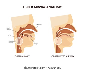
The-anatomy of the upper-airway (Kuna & Remmers, 2000 ... Download scientific diagram | The-anatomy of the upper-airway (Kuna & Remmers, 2000). Factors, causing UA-obstruction, in-patients with sleep-disordered-breathing, are: (1) Skeletal-factors (Verin ...
Upper respiratory airways - Physiopedia
Alveoli Definition, Location, Anatomy, Function, Diagrams Jun 24, 2017 · Alveolar Epithelium. The one-cell thick walls of the alveoli are composed of two distal airway epithelium cell types (pneumocytes) [7]. Type-1 squamous alveolar epithelial cells: Constituting 95% of the alveolar surface area [8], the type 1 cells are extremely thin and flexible to help in the process of gas diffusion so the oxygen-carbon dioxide exchange can occur between the alveoli and the ...
Upper Respiratory System | Respiratory Anatomy
38 complete the labeling of the diagram of the upper ... Complete the labeling of the diagram of the upper respiratory structures (sagittal section). 2. Two pairs of vocal folds are found in the larynx. Anatomy of the Respiratory System - Chute It is a short inch tube that is located in the throat, inferior to the hyoid bone and tongue and anterior to the esophagus.

Pathophysiology of upper airway collapse | Novel Insights ...
PDF Intubation and Anatomy of the Airway And Anesthesia apparatus ANATOMY OF RESPIRATORY TRACT •Anatomically Respiratory tract is divided into upper and lower tract in relation to vocal cord. Upper: nose, mouth, pharynx, larynx, trachea, and mainstem bronchi. Lower: bronchioles , terminal bronchioles, Respiratory bronchioles , alveolar ducts, alveolar sacs, alveoli.

Acute Upper Airway Obstruction | NEJM
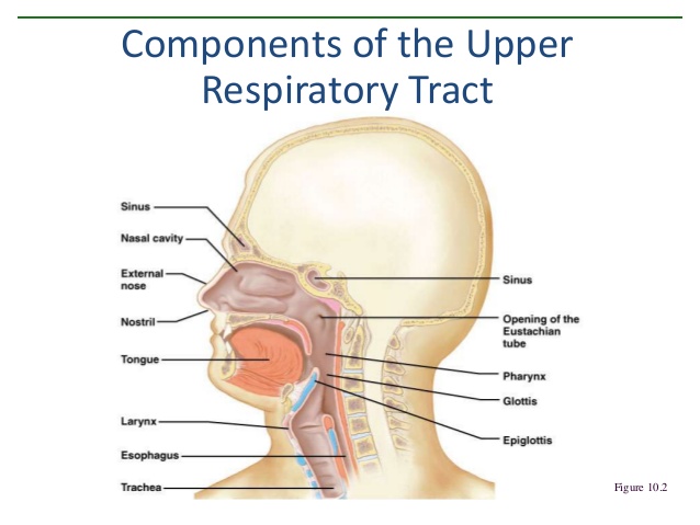
Airway anatomy - SlideShare Airway anatomy. 1. Anatomy of theAnatomy of the Upper AirwayUpper Airway Dr. Saurabh Barde, Anaest hesiology, GMCH, Nagpur. 2. Dr. Saurabh Barde, Anaest hesiology, GMCH, Nagpur. The Upper Airway is a Continuation of the Respiratory System. 3. Components of the Upper AirwAyComponents of the Upper AirwAy NoseNose NasopharynxNasopharynx ...
Anatomy of the Respiratory System in Children | Spectrum ...
Anatomy and physiology of the upper airway An understanding of these structures is important to the clinician involved in maintaining or reestablishing the normal airway. The fol … Anatomy and physiology of the upper airway Anesthesiol Clin North Am. 2002 Dec;20(4):733-45, v. doi: 10.1016/s0889-8537(02)00017-2. ...
What structures are in the upper respiratory tract? and the ...
Anatomy and Physiology of the Upper Airway | Proceedings ... Anatomy and Physiology of the Upper Airway Asli Sahin-Yilmaz 1. x. Asli Sahin-Yilmaz. Search for articles by this author , and Robert M. Naclerio 2. x ... The complaint of alternating nasal obstruction with an acute upper respiratory tract infection shows how the nasal cycle becomes apparent during an illness .

Applied Functional Anatomy of the Airway - Manual of ...
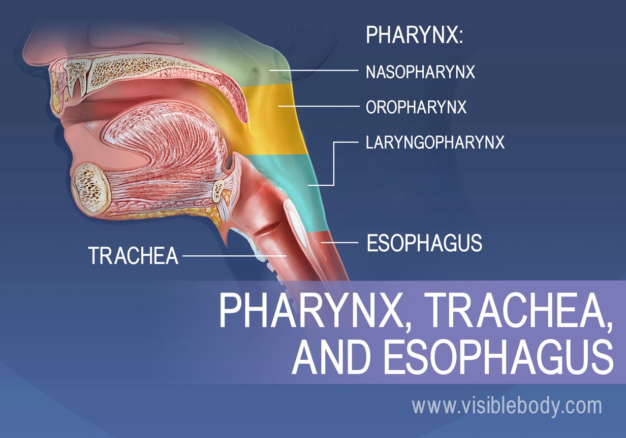
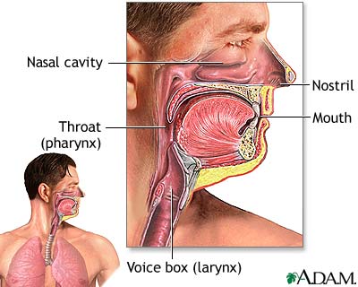
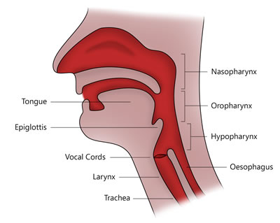
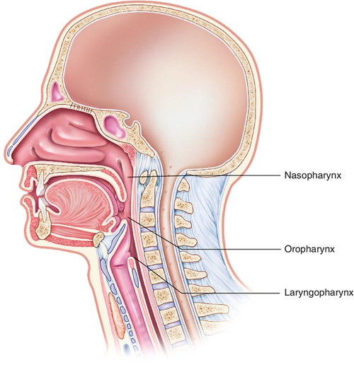
Upper Airway Anatomy - OpenAnesthesia Upper Airway Anatomy Definition The upper airway consists of the pharynx and the nasal cavities; however, some authors include the larynx and trachea as well. The pharynx is can be divided into the nasopharynx, oropharynx, and laryngopharynx. The nose is composed of bone and cartilage, which are in turn attached to the facial skeleton.

RT 127
PPT - Anatomy of the Upper Airway PowerPoint presentation ... Our new CrystalGraphics Chart and Diagram Slides for PowerPoint is a collection of over 1000 impressively designed data-driven chart and editable diagram s guaranteed to impress any audience. They are all artistically enhanced with visually stunning color, shadow and lighting effects. ... "Anatomy of the Upper Airway" is the property of its ...

Anatomy and Physiology of Respiratory System Tutorial
Pharynx: Anatomy & Definition - Video & Lesson Transcript ... Aug 25, 2021 · The nasopharynx is the most superior, or uppermost, part of the pharynx that works with the nasal cavity during breathing. It provides a passageway for air moving from the nose. The pharyngeal ...

Upper respiratory System Quiz
Upper Respiratory Tract: Anatomy, Functions, Diagram Upper Respiratory Tract Structural and Functional Anatomy Nose and Nasal Cavity The nostrils, the two round or oval holes below the external nose, are the primary entrance into the human respiratory system [5]. Lying just after the nostrils are the two nasal cavities, lined with mucous membrane, and tiny hair-like projections called cilia [6].
Airways anatomy Images, Stock Photos & Vectors | Shutterstock
Basic Airway Anatomy - MedicTests The upper airway is the "A" of the ABC's. As the entry point for oxygen any damage to, or blockage of, the structures in the upper airway can rapidly result in unconsciousness or death. The anatomy of the upper airway can be broken down into the nose, mouth, and throat. The medical terms for these are the nasopharynx and oropharynx/larynx.
Anatomy of the Respiratory System | Basicmedical Key
Airway Anatomy - Primary LO of the Day BT_AM 1.1 Describe the anatomy of the upper airway, larynx and trachea … To get the most benefit out of this post, print out the diagram below. Some of the questions need you to draw on it. Name structure A (be specific!) Structure A is the most superior part of the larynx T / F Name structure B On the diagram above, DRAW the following structures:
Upper respiratory tract: MedlinePlus Medical Encyclopedia Image
Anatomy of the Throat and Esophagus - Video & Lesson ... Aug 26, 2021 · The esophagus passes behind the airway to the lungs and in front of the spinal column. It pierces the diaphragm before reaching the stomach. Esophageal muscles move in a wavelike pattern to ...

Respiratory tract - Wikipedia
Amazing Detailed Respiratory System Diagram - Glaucoma ... Air enters the nose through the nostrils. All of the respiratory passages facilitate water and heat exchange which is especially important for dog. Detailed Lungs Diagram Dld02 Lung Anatomy Anatomy Nursing Notes As the names suggest the upper respiratory tract includes everything above the vocal folds furthermore the lower respiratory tract includes everything below the
Upper Airway Anatomy and Function | Ento Key
PDF Airway Anatomy - clemc.us Upper Airway Anatomy Functions warm, filter, and humidify air Nasal cavity and nasopharynx Formed by union of facial bones Nasal floor towards ear not eye Lined with mucous membranes, cilia Tissues are delicate, vascular Adenoids Lymph tissue - filters bacteria Commonly infected Upper Airway Anatomy Oral cavity and oropharynx Teeth Tongue

The Respiratory System - Anatomy & Physiology - The Nursing ...
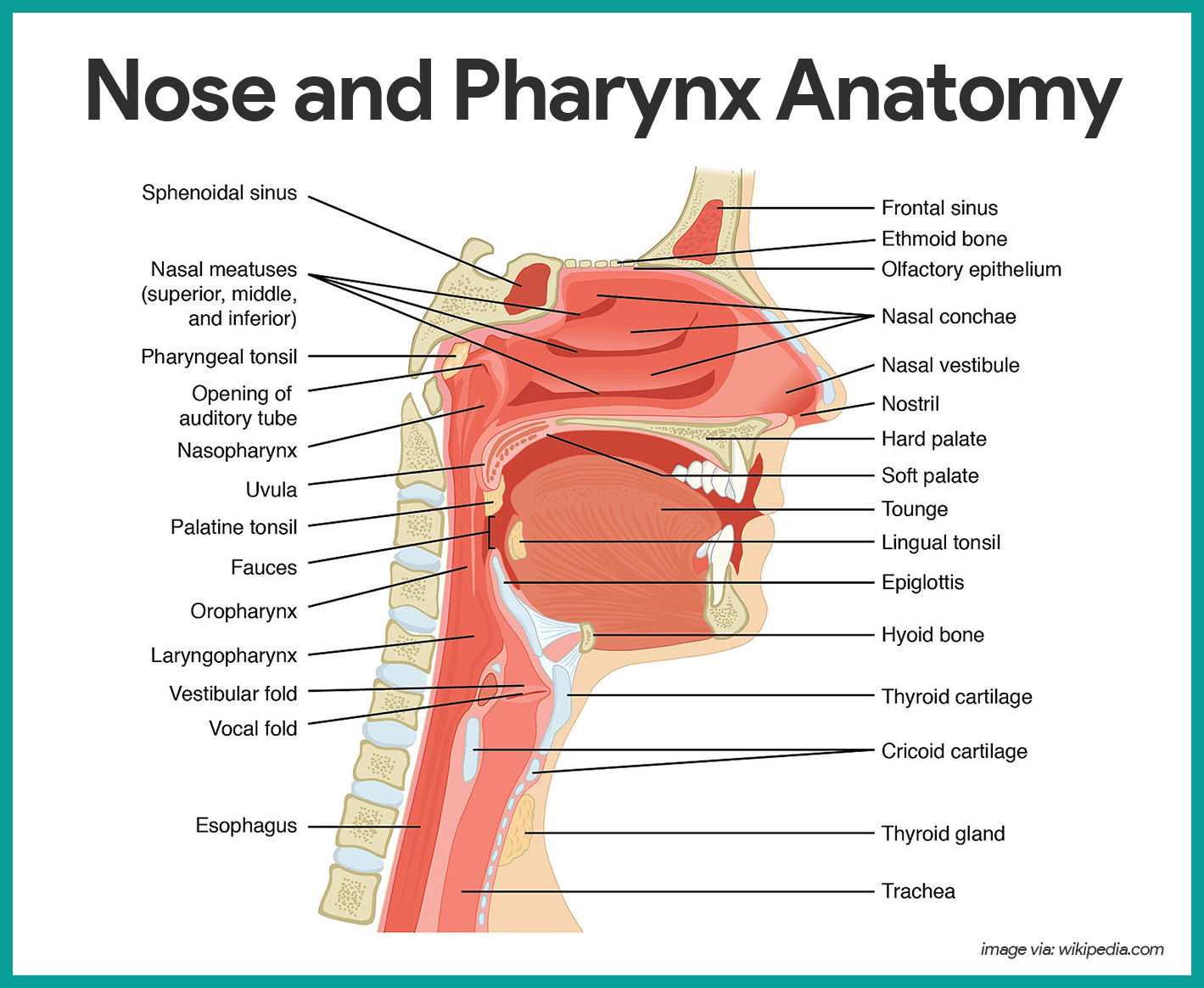
upper airway anatomy Flashcards and Study Sets | Quizlet Upper Airway Anatomy Diagram Sphenoidal Sinus Nasal Meatuses (Superior, middle, and i… Pharyngeal Tonsil ... ... ... 41 terms kayli8 Upper Airway Anatomy 1 Trigeminal Nerve Glossopharynegeal 3 components of upper airway Forms the lingual nerve, provides sensation to anterior 2/3 to… Posterior 1/3 tongue. soft palate, and oropharynx are innervat…

Chapter 22 Respiratory System Part1 - YouTube
Upper Respiratory System | Respiratory Anatomy 4. The Larynx and Vocal Cords Allow Us to Breathe and Talk and Sing The larynx connects the lower part of the pharynx, the laryngopharynx, to the trachea. It keeps the air passages open during breathing and digestion and is the key organ for producing sound. This larynx is comprised of nine cartilages.

Anatomy Of Human Upper Airway While Sleeping With Snoring ...
Anatomy and Physiology: The Upper Respiratory System The nasal cavity is a chamber of the internal nose that connects to the nasopharynx. Air is inhaled through the nostrils and warmed as it passes through the nasal cavities. There is a mucous membrane that lines the cavity; the mucus helps trap unwanted particles in inhaled air. Ethmoid and Other Skeletal Structures
Anatomy of the Respiratory System in Children
PDF 1 Airway Anatomy - Notes For ANZCA Primary Exam 1.Airway Anatomy - 2 Basic Structural Anatomy of Upper Airway & Larynx Laryngeal Skeleton • Hyoid: o Attachement for thyroid cartilage via throhyoid membrane & extrinsinc mm of larynx o U shaped body o Body & paired greater & lesser horns • Skeleton has 9 cartilges: o Singles: ! Thyroid ! Cricoid

Anatomy of a Child's Lung - Pediatric Pulmonologists
EMT Anatomy - Airway Diagram | Quizlet Start studying EMT Anatomy - Airway. Learn vocabulary, terms, and more with flashcards, games, and other study tools.
Anatomy and Physiology (A&P) of the Upper Airway | First Aid ...
Respiratory System Anatomy - Major Zones & Divisions

Upper respiratory airways - Physiopedia
Anatomy of the Respiratory System
8. Anatomy and Physiology of Respiration and Airway ...
Respiratory System - Physiopedia

Anatomy of the upper airway showing the main segments ...

Respiratory System, Anatomy of Your Child's

Continuing Professional Development: Vital airway management ...
Blood and the respiratory system - OpenLearn - Open University

Upper Airway Anatomy Diagram | Quizlet
The Anatomy and Physiology of the Respiratory System. - ppt ...

Anatomy of the upper airway and tongue. (A) Sagittal section ...
Upper respiratory airways - Physiopedia
Upper Respiratory System Images – Browse 1,950 Stock Photos ...
Respiratory System Anatomy and Physiology - Nurseslabs
Anatomy of Upper Airway Stock Photo - Alamy
0 Response to "38 upper airway anatomy diagram"
Post a Comment