41 gram-negative bacteria diagram
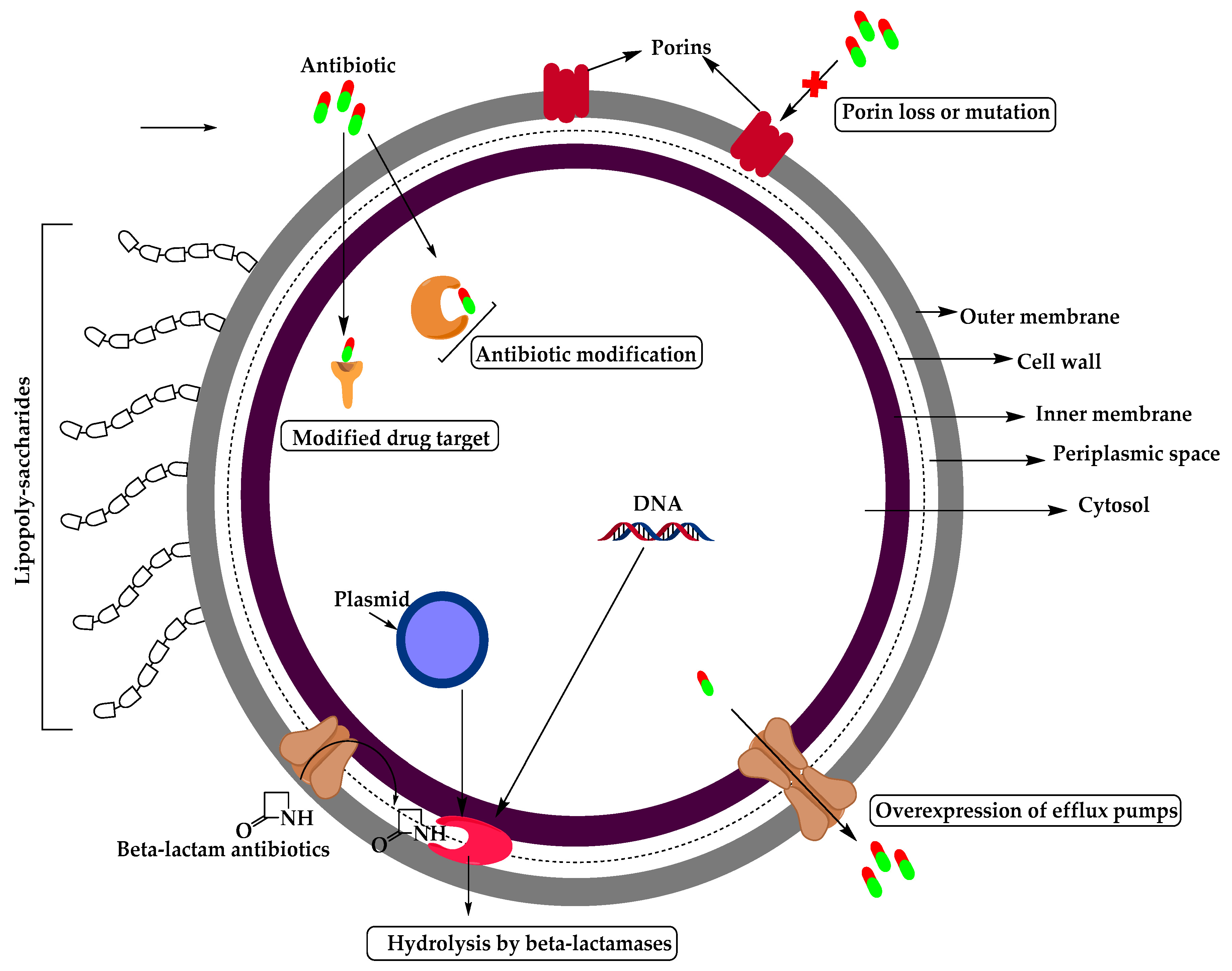
Molecular Expressions Cell Biology: Bacteria Cell Structure Nov 13, 2015 · In gram-negative bacteria, the cell wall is thin and releases the dye readily when washed with an alcohol or acetone solution. Cytoplasm - The cytoplasm, or protoplasm, of bacterial cells is where the functions for cell growth, metabolism, and replication are carried out. Gram-negative Bacteria: Characteristics, List, Cell wall ... Antibiotics for gram-negative bacteria. Gram-negative bacteria are mostly associated with severe, life-threatening infections. These can involve the bloodstream, surgical site, urinary tract, lungs, etc. Despite the use of antibiotics, gram-negative infections are commonly associated with high morbidity and mortality.
Dichotomous Key for Gram Negative Bacteria [classic ... Dichotomous Key for Gram Negative Bacteria [classic] Use Creately's easy online diagram editor to edit this diagram, collaborate with others and export results to multiple image formats. You can edit this template and create your own diagram. Creately diagrams can be exported and added to Word, PPT (powerpoint), Excel, Visio or any other ...

Gram-negative bacteria diagram
Gram negative bacteria examples, list, and structure ... Gram negative bacteria include some bacterial cell examples that possess a thin peptidoglycan cell wall structure. They can cause diseases in humans and stain pink in a gram stain test. In other words, gram-negative bacteria can be defined as the bacterial cells that do not take up the crystal violet stain used in the Gram staining procedure and therefore stain pink at the end of the procedure. CASE STUDY BIOLOGY.docx - Running head: BIOLOGY 1 ... BIOLOGY 2 Extr case study I 1. A gram-negative reaction From the diagram, the bacterial appears to be reddish, which shows it did not retain the crystal violet. In contrast, in gram staining, a differential stain was done to differentiate between gram-negative and gram-positive bacteria and distinguish their morphological appearance. The reaction of the bacteria in the diagram is, therefore, a ... Bacteria - Definition, Shapes, Characteristics, Types & Examples Oct 04, 2019 · This diagram depicts the numerous shapes of bacteria. Bacterial morphology diagram Types of Bacteria. The cell wall also makes Gram staining possible. Gram staining is a method of staining bacteria involving crystal violet dye, iodine, and the counterstain safranin.
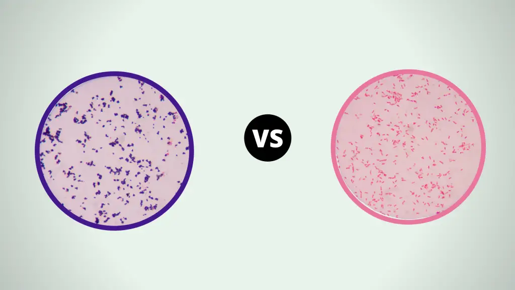
Gram-negative bacteria diagram. Gram Negative Bacterial images | Download Scientific Diagram Gram positive bacterial remains constant with purple or blue color and Gram negative bacteria accomplishes pink or red color due to counter staining. The staining procedure is shown in Figure1. The surface lipoproteins of gram-negative bacteria ... Schematic diagram of SLP vaccine development pipeline. Bioinformatics and reverse vaccinology can be used to identify candidate SLPs. This can be facilitated by searching for translocation machinery such as Slams. Pictured is a pruned phylogenetic tree (see text for details) of pathogenic gram-negative bacteria. Peptidoglycan Function & Structure | What is Peptidoglycan ... A diagram showing the difference between the cell envelope of gram-positive and gram-negative bacteria. Peptidoglycan Functions Peptidoglycan functions for bacteria are numerous. Bacteria shapes, structure and diagram - Jotscroll After that, they are again stained using a certain dye and then viewed with a light microscope. This procedure is called Gram staining. When the bacteria retain the color of the stain, it is called Gram-negative bacteria; when the bacteria fail to retain the color of the stain (becomes violet) it is called Gram-positive bacteria.
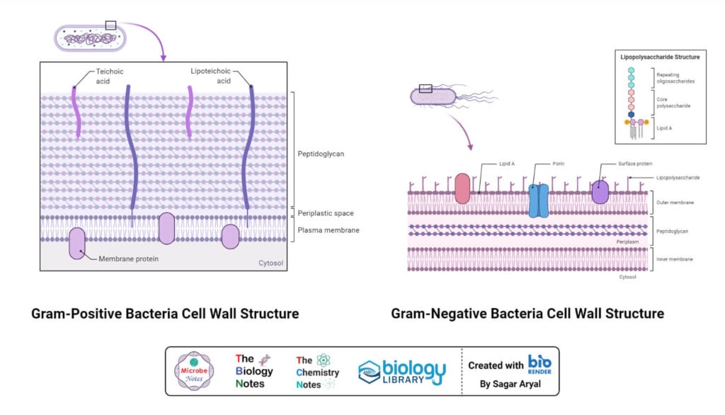
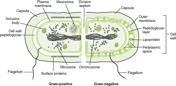
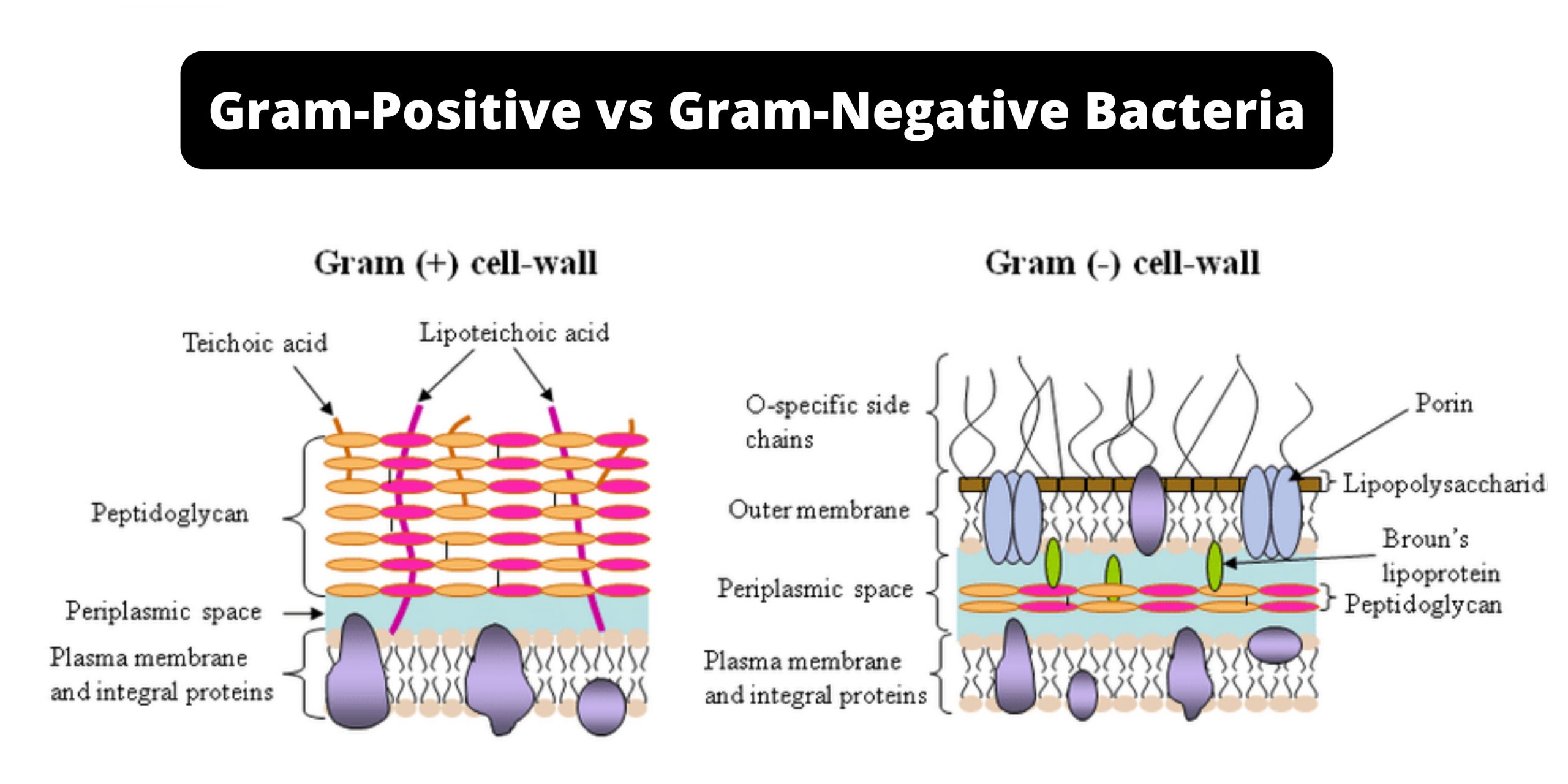
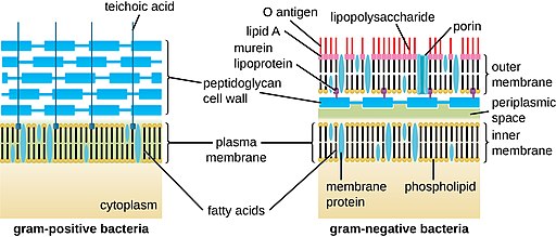
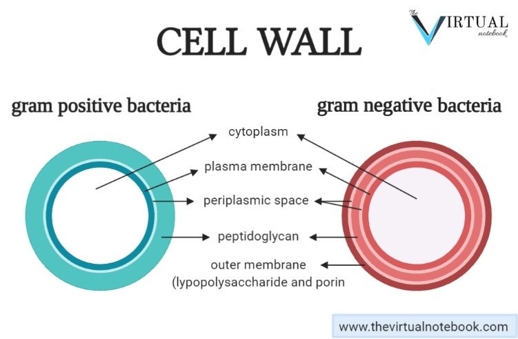
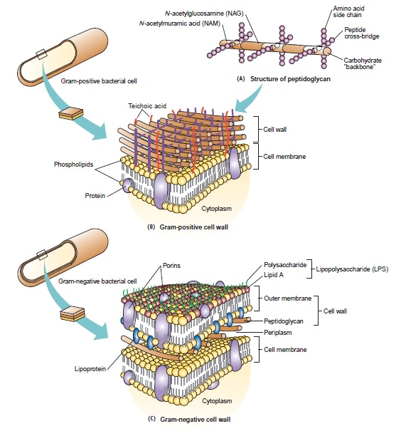
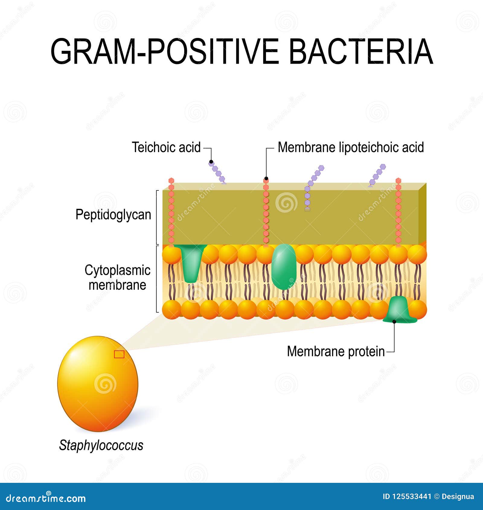
Structure of Bacteria (With Diagram) | Microbiology 8. According to Peberdy (1980) the only compound present in the cell walls of both Gram-negative and Gram-positive bacteria is 'peptidoglycan'. The cell walls of Gram-positive bacteria contain up to 95% peptidoglycan and up to 10% teichoic acids. 9. Cytoplasmic membrane is a thin (5-10 nm) layer lining the inner surface of the cell wall. Gram Positive vs Gram Negative | Technology Networks The diagram below illustrates the differences in the structure of Gram positive and Gram negative bacteria. The two key features that lead to the differing visualization properties of Gram positive and Gram negative species are the thickness of the peptidoglycan layer and presence or absence of the outer lipid membrane. Gram Positive and Gram Negative Bacteria - Structures ... As with Gram positive bacteria, Gram negative bacteria also contain the peptidoglycan polymer in their cell wall. While this polymer is thin (2 to 4 nanometers in thickness with just about 3 layers of peptidoglycan) in Gram negative bacteria, it's also composed of long glycan strands that are cross-linked by peptide molecules. Gram Positive Bacteria - Gram Project Gram positive bacteria types and classification. Gram Project is a medical education resource website containing diagrams, tables and flowcharts for all your quick referencing, revision and teaching needs.
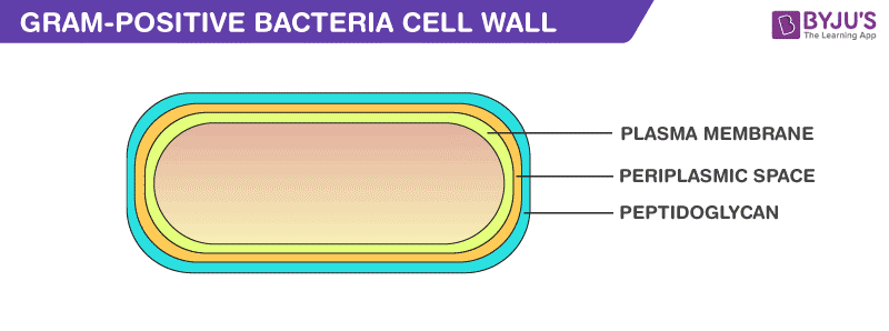
Solved t. Draw aned iabel a agram of a GENERAL bacterial ... Transcribed image text: t. Draw aned iabel a agram of a GENERAL bacterial cell on & on your d o Cell wall Plasma membrane (cell membrane) Flagella o Wnte out short description of the function or characteristics for each labeled component on the drawing. 2 Draw and Label a diagram of the membrane and cell wall structure of Gram Positive and Gram negative bacteria and label the following ... Bacterial Cell Wall Structure: Gram-positive & negative Gram-negative Bacteria. Go to PAGE 2 > Gram-positive Cells. In Gram-positive bacteria, peptidoglycan makes up as much as 90% of the thick cell wall enclosing the plasma membrane. See Page 2 for a diagram of the Gram-negative cell wall and a video on ... Difference Between Gram-positive and Gram-negative Bacteria The gram-positive bacteria retain the crystal violet colour and stains purple whereas the gram-negative bacteria lose crystal violet and stain red. Thus, the two types of bacteria are distinguished by gram staining. Gram-negative bacteria are more resistant against antibodies because their cell wall is impenetrable. Gram negative diplococci bacteria: Introduction ... Gram Staining: The diagnosis is suggested by the finding of gram-negative bacteria bean-shaped capsular ( mark of evidence in Gram stain) diplococci as shown above image but the capsular mark is not recovered gram stain from culture. Culture: The organism is cultured on blood agar or chocolate agar incubated at 37°C in a 5% CO 2 atmosphere.
Gram-negative bacteria - Wikipedia
Electron Transport Chain of Bacteria (With Diagram) ADVERTISEMENTS: The electron transport chains of bacteria (prokaryotes) operate in plasma membrane (mitochondria are absent in prokaryotes). Some bacterial electron transport chains resemble the mitochondrial electron transport chain. Paracoccus denitrificans is a gram-negative, facultative anaerobic soil bacterium. It is a model prokaryote for studies of respiration. When this bacterium grows ...

Through the wall: extracellular vesicles in Gram-positive ...
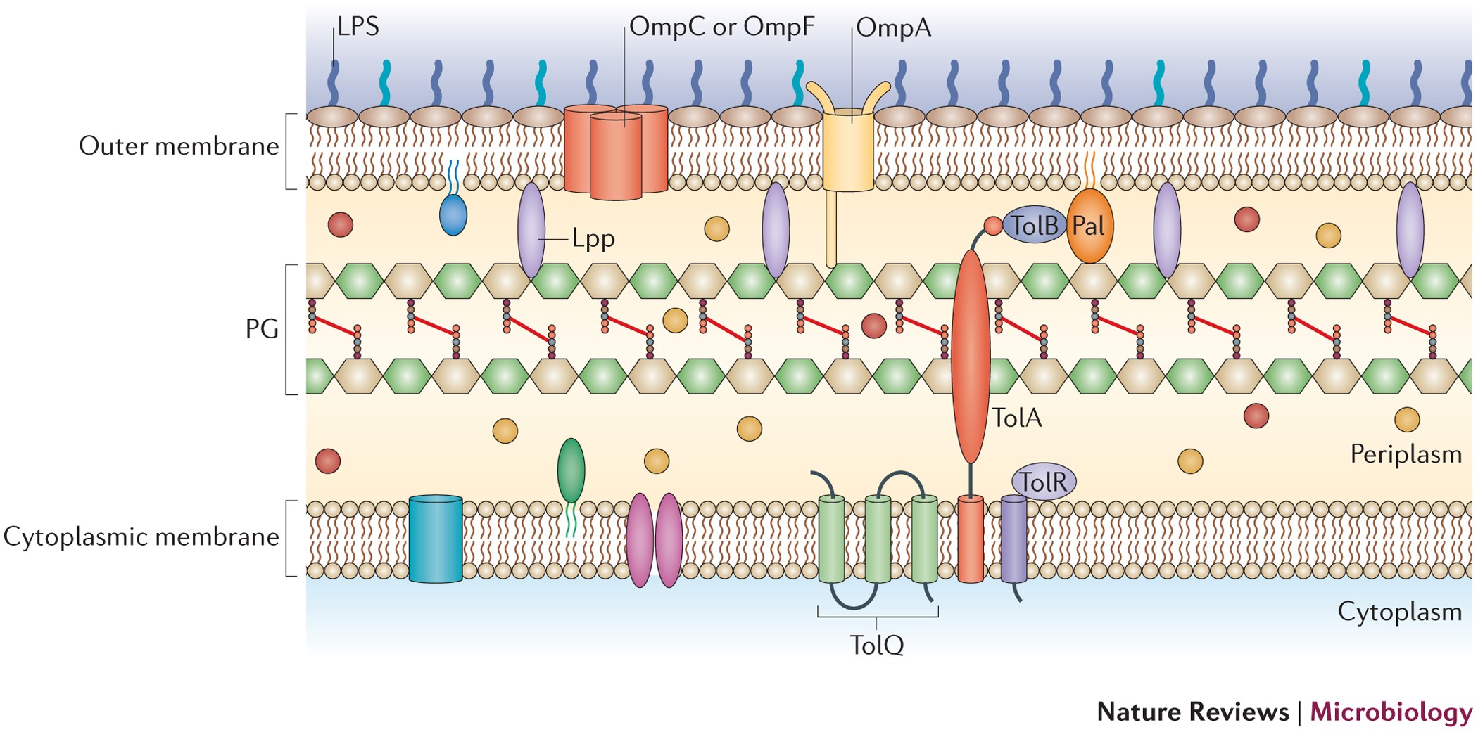
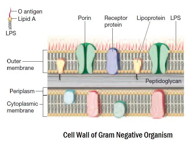
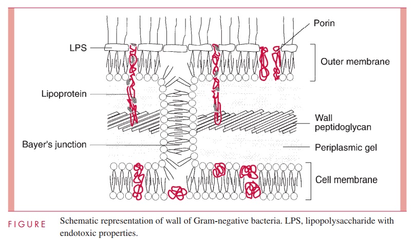
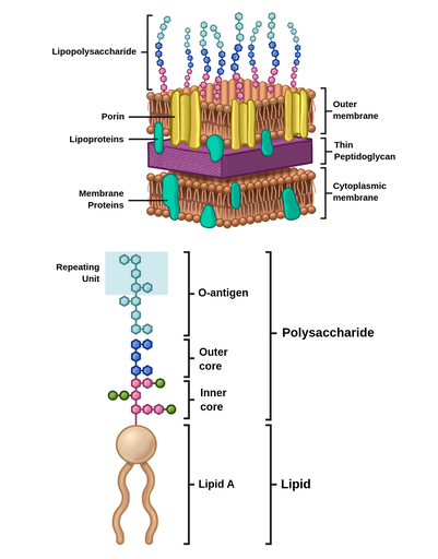
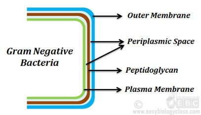
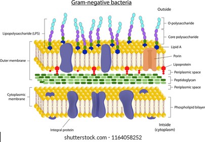
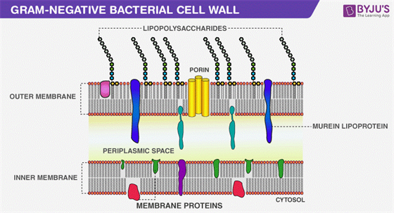
Structure of Gram-negative cell wall Schematic diagram of the cell wall of the Gram-negative bacteria. Outer membrane The membrane's outer layer has a two-layered design. its inner layer is similar the composition of the membrane of cells, and the outer membrane is made up of a distinct component known as lipopolysaccharide.
30 Comparison between Gram-Positive and Gram-Negative Bacteria
Bacteria - Definition, Structure, Diagram, Classification May 17, 2021 · Today, bacteria are considered as one of the oldest forms of life on earth. Even though most bacteria make us ill, they have a long-term, mutual relationship with humans and are very much important for our survival. But before we elaborate on its uses, let us know the structure of bacteria, its classification, and the bacteria diagram in detail.
Gram Staining- Principle, Reagents, Procedure, Steps, Results
Gram Negative Bacteria Flow Chart - Reviews Of Chart Solved Gram Positive Flow Chart The Graphic Below Is Able Move Your Mouse Over An Item On And If Arrow Turns Into A Hand Cl Course Hero. Gram Negative Flowchart Docx Bacteria Quynh Dinh Oxidase Test Mr Urease Enterobacter Aerogenes Course Hero. Flow diagram of bloodstream infection gram negative bacterial isolates scientific dichotomous key ...
Outer-membrane vesicles from Gram-negative bacteria ...
Gram-Negative Bacilli: Characteristics, Types & Examples ... Jan 21, 2022 · Gram-negative bacilli are several types of bacteria based on their shape and stain color when viewed under a microscope. Discover the characteristics defining these bacteria, the ...

bacteria_101_cell_walls_gram_staining_common_pathogens [TUSOM ...
Structures of Gram-Negative Cell Walls and Their Derived ... Gram-negative cell walls are strong enough to withstand ∼3 atm of turgor pressure (), tough enough to endure extreme temperatures and pHs (e.g., Thiobacillus ferrooxidans grows at a pH of ≈1.5) and elastic enough to be capable of expanding several times their normal surface area ().Strong, tough, and elastic … the gram-negative cell wall is a remarkable structure which protects the ...

Endotoxin Gram-negative bacteria Gram-positive bacteria ...
Gram-Negative Bacteria | List, Characteristics & Types ... Gram-negative rods are bacteria which have a rod, or bacillus, shape, and are also gram negative as a result of having a thin layer of peptidoglycan and an outer membrane. Due to their rod shape,...

Bacteria basics – Part 2 – ReAct
Gram-negative bacteria- cell wall, examples, diseases ... The cell wall of gram-negative bacteria is complex having a thin layer of the peptidoglycan layer of 2-7nm and a thick outer membrane of 7-8nm thick. Microscopically, there is a space that is seen between the cell membrane and the cell wall, known as the periplasmic space made up of periplasm.

Bacterial Cell Wall Structure: Gram Positive vs Gram Negative
Gram-negative bacteria - Wikipedia Gram-negative bacteria are bacteria that do not retain the crystal violet stain used in the Gram staining method of bacterial differentiation. They are characterized by their cell envelopes, which are composed of a thin peptidoglycan cell wall sandwiched between an inner cytoplasmic cell membrane and a bacterial outer membrane .
Differences Between Gram Positive and Gram Negative Bacteria ...
Percentages of Gram positive and gram negative bacteria ... Download scientific diagram | Percentages of Gram positive and gram negative bacteria. Total number of Gram positive bacterial colonies = 15 (36%) Total number of Gram negative bacterial colonies ...
Bacterial Structure | Clinical Gate
Spirilla Bacteria - Definition, Examples, Shape, Diseases Like all the other Spirilla bacteria, all members of this group are Gram-negative. They are also mostly microaerophilic and thus require a low level of oxygen to survive. However, based on culture techniques, studies have shown that they can grow aerobically in certain liquid media (media with special supplements).

Gram negative bacteria examples, list, and structure - Jotscroll
Gram Negative Bacteria - Gram Project - Medical diagrams ... Gram negative bacteria types and classification flowchart. Gram Project is a medical education resource website containing diagrams, tables and flowcharts for all your quick referencing, revision and teaching needs.
Describe the structure of Gram positive and Gram negative ...
Gram Positive and Gram Negative Bacteria Diagram | Quizlet Start studying Gram Positive and Gram Negative Bacteria. Learn vocabulary, terms, and more with flashcards, games, and other study tools.

Gram Positive vs Gram Negative | Technology Networks
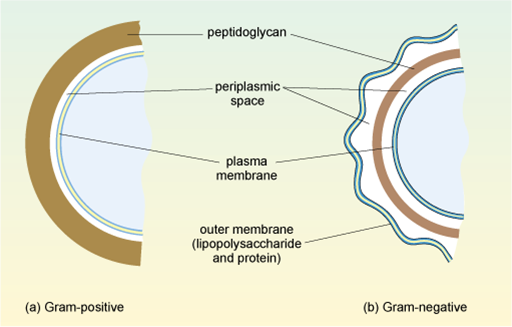
Structures of Gram‐Positive and Gram‐Negative Bacterial ... Gram-negative bacteria have a smaller amount of this rigid structure than do gram-positive bacteria. Periplasmic space. an open area between the cell wall and cell membrane in the cell envelopes of bacteria. gram negative bacteria have a more extensive space than do a gram positive bacteria. Cell membrane.
Difference between Gram-Positive and Negative Bacteria - Gram ...
Interactive Bacteria Cell Model - CELLS alive Mycoplasma are bacteria that have no cell wall and therefore have no definite shape. Outer Membrane: This lipid bilayer is found in Gram negative bacteria and is the location of lipopolysaccharide (LPS) in these bacteria. Gram positive bacteria lack this layer. LPS can be toxic to a host and can stimulate the host's immune system.

6. Structure of Gram-positive and Gram-negative bacteria ...
Gram Positive vs. Gram Negative Bacteria - ThoughtCo The cell wall structure of Gram negative bacteria is more complex than that of Gram positive bacteria. Located between the plasma membrane and the thin peptidoglycan layer is a gel-like matrix called periplasmic space. Unlike in Gram positive bacteria, Gram negative bacteria have an outer membrane layer that is external to the peptidoglycan ...

Difference between Gram Positive and Gram Negative Bacteria
Bacteria - Definition, Shapes, Characteristics, Types & Examples Oct 04, 2019 · This diagram depicts the numerous shapes of bacteria. Bacterial morphology diagram Types of Bacteria. The cell wall also makes Gram staining possible. Gram staining is a method of staining bacteria involving crystal violet dye, iodine, and the counterstain safranin.
Gram Negative Cell Wall - Bacterial Structures
CASE STUDY BIOLOGY.docx - Running head: BIOLOGY 1 ... BIOLOGY 2 Extr case study I 1. A gram-negative reaction From the diagram, the bacterial appears to be reddish, which shows it did not retain the crystal violet. In contrast, in gram staining, a differential stain was done to differentiate between gram-negative and gram-positive bacteria and distinguish their morphological appearance. The reaction of the bacteria in the diagram is, therefore, a ...
Gram-negative cell wall - Labster Theory
Gram negative bacteria examples, list, and structure ... Gram negative bacteria include some bacterial cell examples that possess a thin peptidoglycan cell wall structure. They can cause diseases in humans and stain pink in a gram stain test. In other words, gram-negative bacteria can be defined as the bacterial cells that do not take up the crystal violet stain used in the Gram staining procedure and therefore stain pink at the end of the procedure.

Structure and Function of Bacterial Cells
Gram Positive Vs Gram Negative Bacteria: A Comparison | Easy ...
Gram-negative Images, Stock Photos & Vectors | Shutterstock
Molecules | Free Full-Text | Resistance of Gram-Negative ...
Cell Wall - Gram Positive vs Negative Bacteria | Easy Biology ...

Bacteria cell wall gram positive and gram negative

4: Bacteria - Cell Walls - Biology LibreTexts
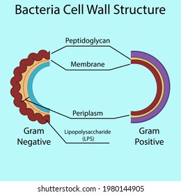
Diagram Gram Negative Gram Positive Bacteria Stock Vector ...

Gram Positive vs Gram Negative | Technology Networks
Gram negative bacteria, cell wall, diseases - The Virtual ...
Understanding antibiotic resistance - OpenLearn - Open University

Cell Wall Structure Gramnegative Bacteria Example Stock ...
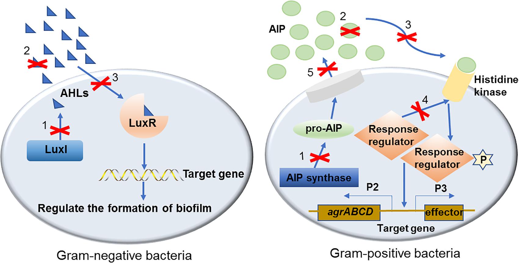
Frontiers | Regulatory Mechanisms and Promising Applications ...

Electrochemical cell lysis of gram-positive and gram-negative ...
Print page
Gram Positive Bacteria - Characteristics And Structure
Cell Wall Structure of Gram-positive Bacteria for Example ...
How can we change gram negative to gram positive in bacteria ...

Surface Proteins of Gram-Positive Bacteria and Mechanisms of ...

Gram-Positive vs. Gram-Negative Bacteria • Microbe Online

Microbiological educational diagram sample: Cell envelope of ...
Gram Negative Bacteria - Characteristics, Cell Structure and ...
0 Response to "41 gram-negative bacteria diagram"
Post a Comment