39 mitochondria diagram with labels
Botany - Biology Discussion Usually, mitochondria are 0.5 to 1 n in diameter and 3-6n in length. They, however, vary in their size and are also capable of changing their size. 4. They are generally rod shaped but may be in the form of granules or spherical bodies. 5. Their number may vary from 50 to 50,000 in different kinds of cells. ADVERTISEMENTS: 6. Plant Cell Diagram Mitochondria Labeled The mitochondrion (/ ˌ m aɪ t ə ˈ k ɒ n d r ɪ ə n /, plural mitochondria) is a double membrane-bound organelle found in most eukaryotic organisms. Where are Mitochondria Located They are found in the cytoplasm of nearly all plant and animal cells. Muscle cells need a lot of energy so they have loads of mitochondria.
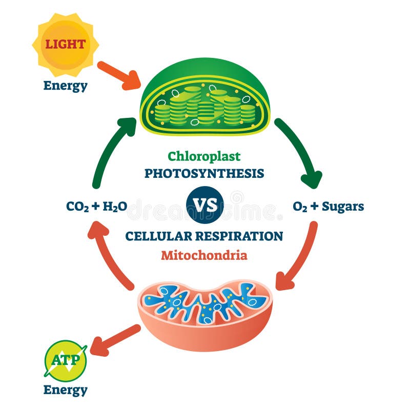
Draw labelled diagram and describe the structure and ... OR Draw a well labelled diagram of mitochondria. asked Nov 19, 2020 in Biology by Saavya (51.8k points) cell unit of life; class-11 +1 vote. 1 answer. The three boxes in this diagram represent the three major biosynthetic pathways in aerobic respiration. Arrows represent net reactants or products.
Mitochondria diagram with labels
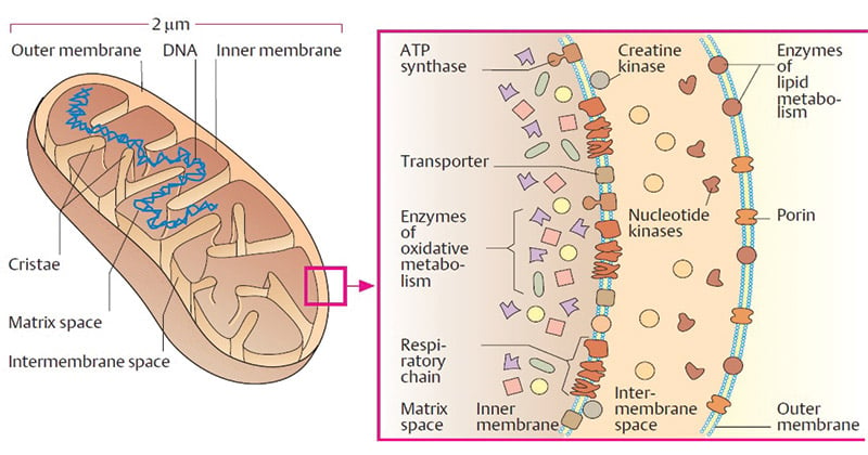
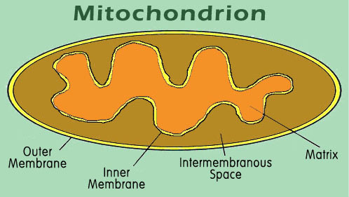
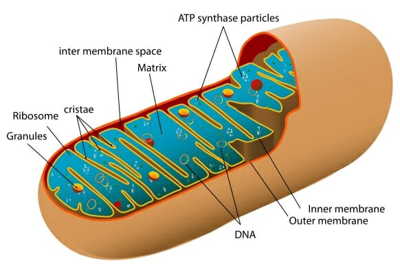
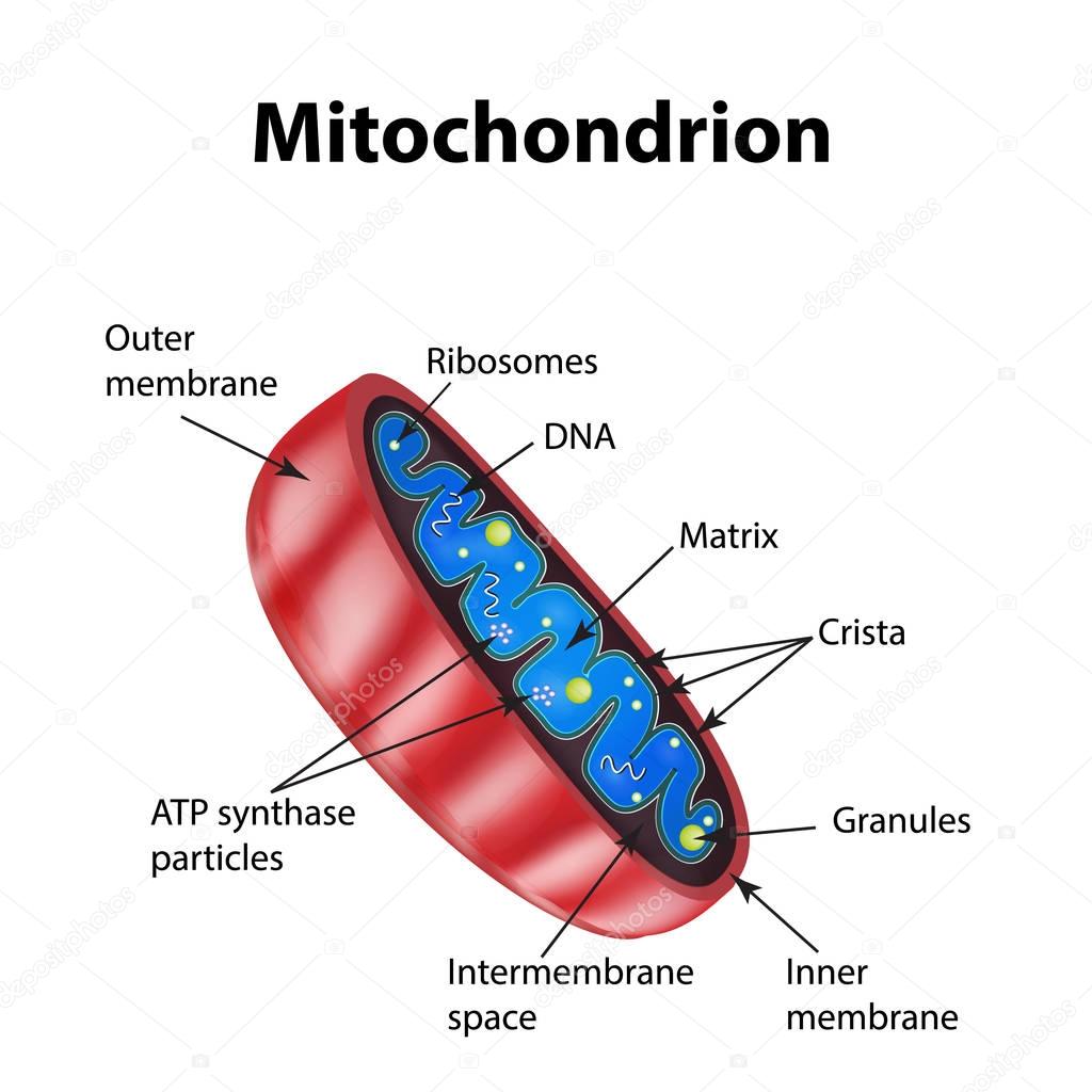
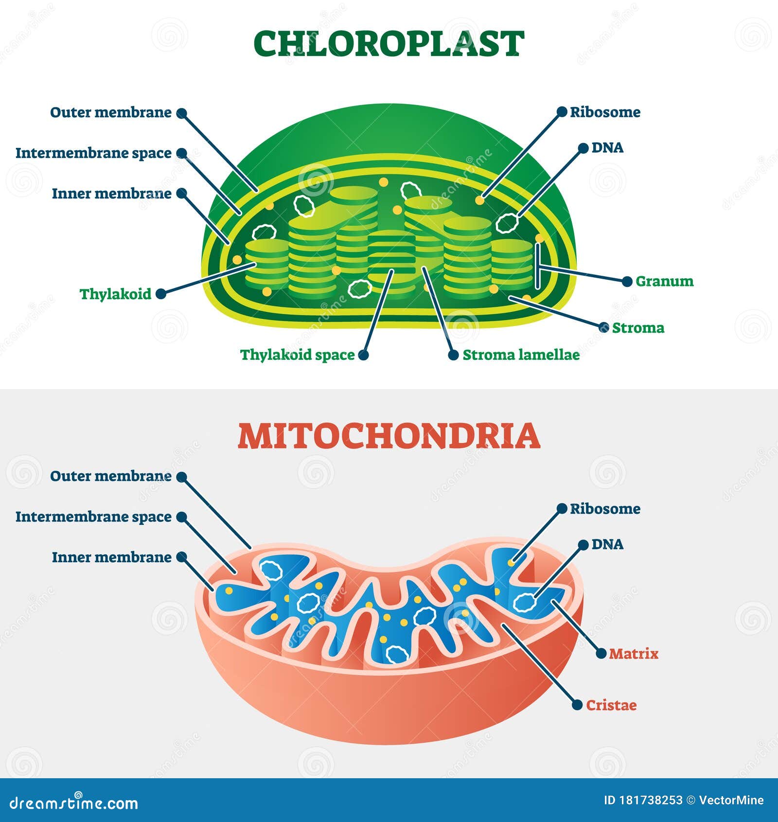
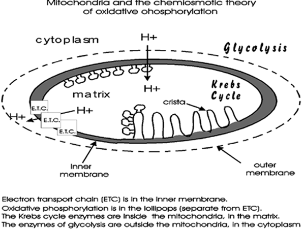
Cytoskeleton Diagram Labeled (B) Mitochondria Labeled With Dsred2. In cell biology the cytoskeleton is a system of fibrillar structures that pervades the cytoplasm. Animal cell diagram labeled cytoskeleton. Microtubules, microfilaments (actin filaments), and intermediate filaments. It Assists In Cell Signalling. The diagram is schematic representation of ... - Brainly.com The diagram is schematic representation of the electron transport mechanism in mitochondria. Label the diagram to complete the model. 2 See answers Advertisement Advertisement smilodon smilodon As shown in the presented diagram in the process of electron transport happens in the mitochondrial matrix. Diagram Of Mitochondria - BYJUS Diagram Of Mitochondria The diagram below shows the structure and functions of the mitochondria. Structure and Functions Of Mitochondria Matrix It is a viscous or a gel-like fluid containing a mixture of enzymes, ribosomes, inorganic ions, mitochondrial DNA, nucleotide cofactors, and organic molecules.
Mitochondria diagram with labels. The diagram is schematic representation of the electron ... The diagram is schematic representation of the electron transport mechanism in mitochondria. Label the diagram to complete the model. 2 See answers Advertisement Advertisement river2738 river2738 The big blue area is the mitochondrial matrix. This is where the citric acid cycle takes place; also known as the Krebs cycle or Tricarboxylic acid cycle. Respiration & Fermentation Review (Questions ... - Course Hero View Respiration & Fermentation Review (Questions & Diagram Label).pdf from BIOLOGY 101 at Jack Britt High School. Name: _ Pd: _ Date: _ Cellular Respiration & Fermentation Review Directions: Using byjus.com › biology › diagram-of-neuronA Labelled Diagram Of Neuron with Detailed Explanations The diagram or the structure of the Neuron is useful for both Class 11 and 12 board exams as it has been repetitively asked in the board examinations. It is also one among the few topics having the highest weightage of marks. Learn More: Difference between Sensory and Motor Neuron. Diagram Of Neuron with Labels Mitochondrial Labeling - Thermo Fisher Scientific Labeling functioning mitochondria Mitochondria are organelles found in every eukaryotic cell. Because they generate ATP, they can be thought of as the power plants of the cell. In addition to energy production, mitochondria also play critical roles in cell signaling, cell cycle, calcium buffering, and both cell growth and death.
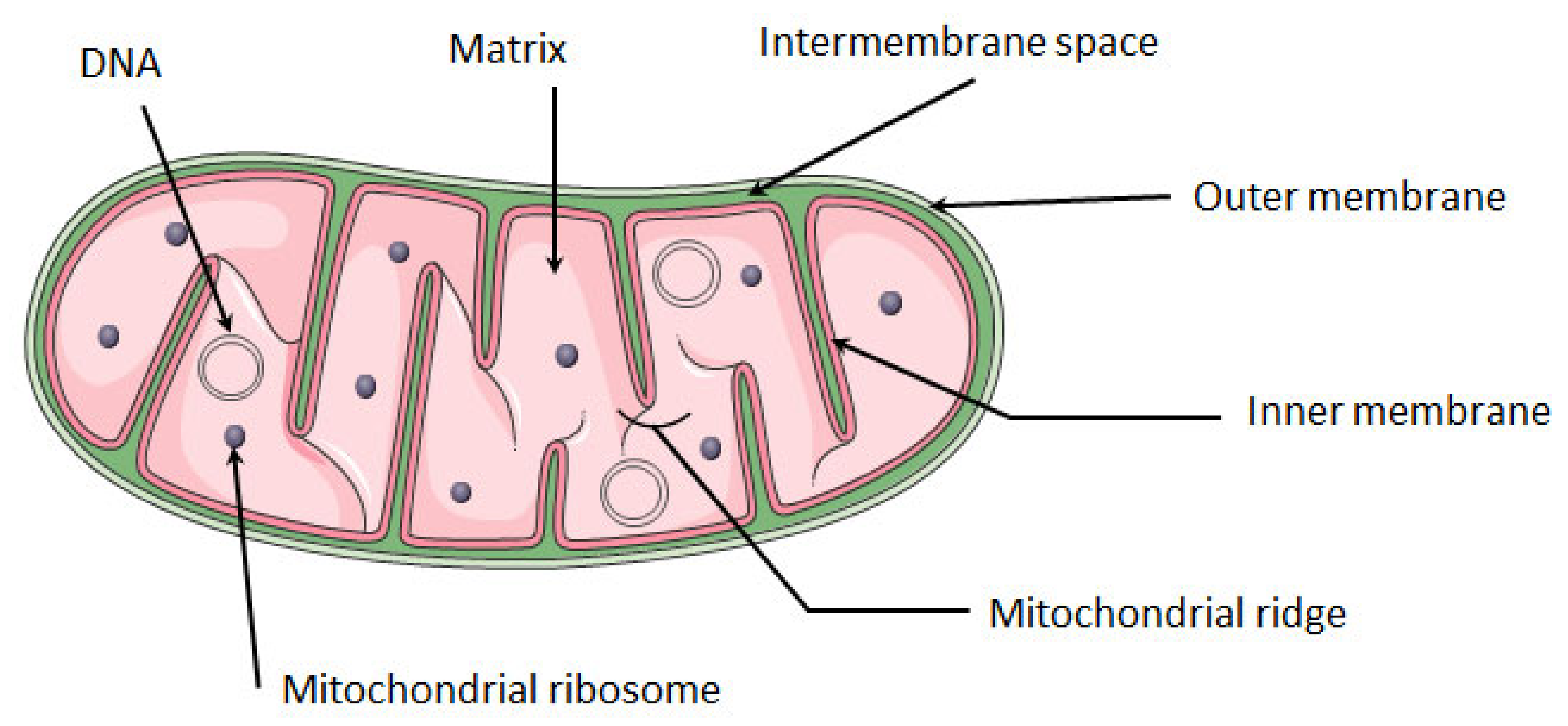
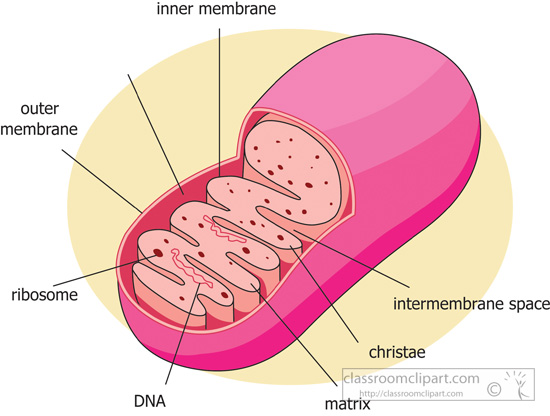
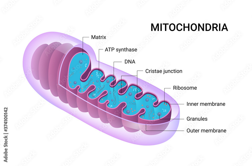
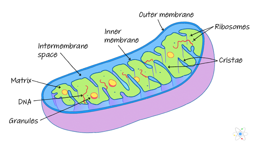
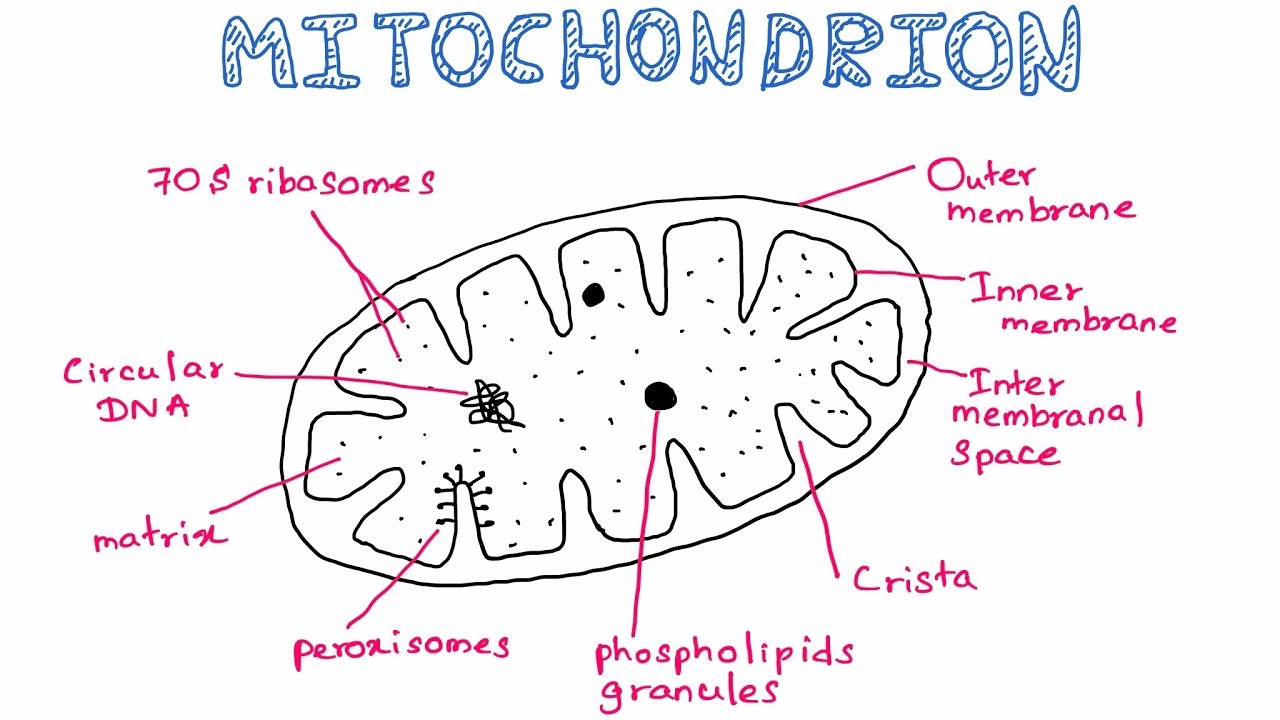
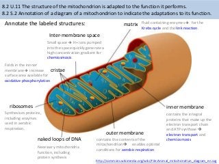
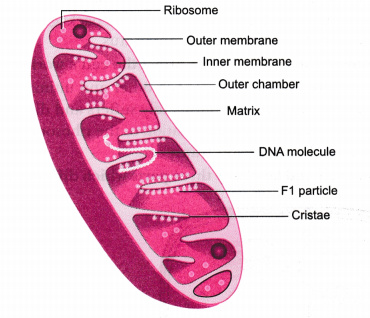
› proteins › proteinProtein Targeting (With Diagram) | Molecular Biology Targeting of Proteins to Mitochondria and Chloroplasts 8. Protein Targeting to Chloroplasts 9. Protein Targeting into Nucleus and 10. Membrane Proteins. Introduction to Protein Targeting: A typical mammalian cell may contain numerous kinds of proteins and numerous individual protein molecules. The eukaryotic cell is a multi-compartmental structure. DOC Read, Answer, Color, Label: Mitochondria The mitochondria are about the size of a bacterial cell and are often peanut-shaped. Mitochondria have their own DNAand a double membranelike the nucleus and the chloroplast. The outer membraneis smooth, while the inner membranehas many folds called cristaewhich help to increase the surface area of the membrane. Energy Production in the Mitochondria Label the substances ... Energy Production in the Mitochondria Label the substances and processes involved in energy production in the mitochondria. Drag the labels onto the diagram to identify the substances and processes involved in energy production in the mitochondria. Animal Cell Diagram Mitochondria Labeled The mitochondrion (plural mitochondria) is a membrane-bound organelle found in the cytoplasm of eukaryotic cells. One of the most intricate tasks that healthiness experts face throughout their interplay with patients helps them realise the problems and the way to motivate them concerning the diagnosis and treatment available.
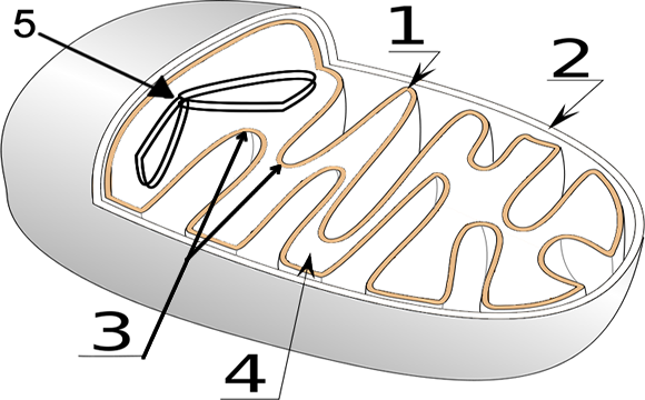
Match the letter in the diagram of a mitochondria Match the letter in the diagram of a mitochondria with the label: (you may use each letter more than once or not at all) Unit 4-6: Photosynthesis -Photophosphorylation and the Calvin Cycle Unit 4-6 Exam Type Questions: 1. In photophosphorylation: A. light is oxidized. B. light is reduced. C. light is the electron source. Cell Organelles- Definition, Structure ... - Microbe Notes Cell Organelles Definition. Cell organelles are specialized entities present inside a particular type of cell that performs a specific function. There are various cell organelles, out of which, some are common in most types of cells like cell membranes, nucleus, and cytoplasm. However, some organelles are specific to one particular type of cell ... quizlet.com › 509259114 › ap-biology-unit-4-chapterAP Biology Unit 4 Chapter 12 Flashcards - Quizlet Drag the labels to their appropriate locations in the table below. The number at the top of each column corresponds to the same number in the image above. Each column describes what happens at that numbered stage. Use only white labels for white targets, blue labels for blue targets, and pink labels for pink targets. quizlet.com › 568033559 › botany-exam-1-chs-1/2/3-4Botany Exam 1 Chs. 1, 2, 3, 4, 5, 6, 7, 12, and 16 Quizzes ... Mitosis, or the division of a mother cell's nucleus into two identical daughter nuclei, is typically divided into four phases. Match each of the labels to identify what events take place during each phase of mitosis. 1. Prophase 2. Metaphase 3. Anaphase 4. Telophase A) Chromosomes are aligned at the equator of the cell and the spindle is fully ...
IJMS | Free Full-Text | Mitochondria as a Cellular Hub in ...
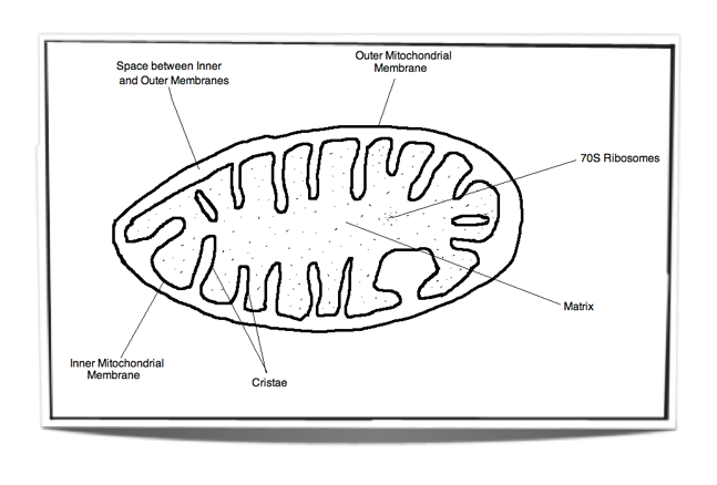
Mitochondria Labeling Diagram - Quizlet The electron transport chain and chemiosmosis takes place on this membrane as part of cellular respiration to create ATP. The cristae increase the surface area of the inner membrane, allowing for faster production of ATP because there are more places to perform the process.

The figure below shows the structure of a mitochondrion with ...
How to label a diagram - BioRender Description: In this short tutorial, learn how to effectively and elegantly label your diagrams. Summary: When you're labelling a diagram: make sure your labels are evenly spaced (0:22) and use parallel label lines with a dot (0:37).Effective label lines create elegantly labelled figures, like anatomy of the brain (3:25). Meet the expert: Shiz Aoki, CEO and co-founder of ...
Diagram of Mitochondria and Function of Mitochondria Parts ...
Mitochondria Labeling Diagram - Quizlet Mitochondria Labeling STUDY Learn Flashcards Write Spell Test PLAY Match Gravity Created by meganplocher734 Terms in this set (5) inner membrane intermembrane space the space inside the mitochondria matrix The syrup of the mitochondria; everything in the inside (Citric Acid Cycle) cristae the folds of the inner membrane (ETC) outer membrane
Mitochondria- Definition, Structure, Functions and Diagram
What Is Mitochondria (Structure, Diagram & Function) - BYJUS Mitochondria diagram explaining the structure of mitochondria Structure of Mitochondria The mitochondrion is a double-membraned, rod-shaped structure found in both plant and animal cell. Its size ranges from 0.5 to 1.0 micrometre in diameter. The structure comprises an outer membrane, an inner membrane, and a gel-like material called the matrix.

Cell anatomy vector illustration. Labeled educational ...
Diagram Of Mitochondria - BYJUS Diagram Of Mitochondria The diagram below shows the structure and functions of the mitochondria. Structure and Functions Of Mitochondria Matrix It is a viscous or a gel-like fluid containing a mixture of enzymes, ribosomes, inorganic ions, mitochondrial DNA, nucleotide cofactors, and organic molecules.
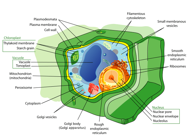
Labeled Plant Cell With Diagrams | Science Trends
The diagram is schematic representation of ... - Brainly.com The diagram is schematic representation of the electron transport mechanism in mitochondria. Label the diagram to complete the model. 2 See answers Advertisement Advertisement smilodon smilodon As shown in the presented diagram in the process of electron transport happens in the mitochondrial matrix.
Shutterstock - PuzzlePix
Cytoskeleton Diagram Labeled (B) Mitochondria Labeled With Dsred2. In cell biology the cytoskeleton is a system of fibrillar structures that pervades the cytoplasm. Animal cell diagram labeled cytoskeleton. Microtubules, microfilaments (actin filaments), and intermediate filaments. It Assists In Cell Signalling.
Science Clipart - mitochondria-diagram-labeled - Classroom ...
Vector diagram of Mitochondria. Cross-section view. Medical ...

toxiology of biochem - Draw a diagram of a typical ...

Mitochondrion structure diagram. Mitochondria have two ...
mitochondria picture with labels - Clip Art Library
Mitochondria: Definition, Structure & Function (with Diagram)
how to draw the mitochondria
8.2 cellular respiration

Mitochondria, Illustration, Labeled Stock Photo - Alamy
Draw labelled diagram and describe the structure and function ...

Mitochondrion - Definition and Examples - Biology Online ...

Printable Animal Cell Diagram – Labeled, Unlabeled, and Blank
Mitochondria - Definition, Function & Structure | Biology ...

Schematic diagram showing different processes of mitochondria ...

How to draw the diagram of mitochondria //easy steps by step ...
Mitochondria Stock Vectors, Royalty Free Mitochondria ...

Animal Cell- Definition, Structure, Parts, Functions, Labeled ...
Chloroplast Vs Mitochondria Vector Illustration. Labeled ...
How Does the Mitochondria Produce Energy for the Cell

Draw a neat diagram and label the following diagram ...
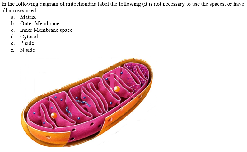
Solved In the following diagram of mitochondria label the ...

how to draw and label mitochondria - YouTube | Biology ...

Mitochondria: Structure, Functions and Diagram – StudiousGuy
Draw a labelled diagram of mitochondria. Write the functions ...
IB Biology Notes - 8.1 Cell respiration

Diagram of a Mitochondrion

Sketch and Label 'Ultrastructure of Mitochondrion ...

Draw a diagram of mitochondria? Label parts how many types of ...
Mitochondria Stock Illustrations – 1,691 Mitochondria Stock ...

Mitochondria Stock Vectors, Royalty Free Mitochondria ...
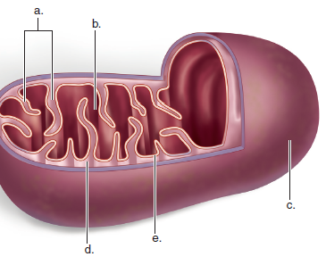
Solved: Label this diagram of a mitochondrion, and state a ...
Mitochondria Handout
0 Response to "39 mitochondria diagram with labels"
Post a Comment